. 2018 Jan 31;15(1):29.
doi: 10.1186/s12974-018-1073-0.

miRNA-23a/CXCR4 regulates neuropathic pain via directly targeting TXNIP/NLRP3 inflammasome axis

Affiliations

miRNA-23a/CXCR4 regulates neuropathic pain via directly targeting TXNIP/NLRP3 inflammasome axis

Zhiqiang Pan et al. J Neuroinflammation. .

Abstract

Background: Chemokine CXC receptor 4 (CXCR4) in spinal glial cells has been implicated in neuropathic pain. However, the regulatory cascades of CXCR4 in neuropathic pain remain elusive. Here, we investigated the functional regulatory role of miRNAs in the pain process and its interplay with CXCR4 and its downstream signaling.

Methods: miRNAs and CXCR4 and its downstream signaling molecules were measured in the spinal cords of mice with sciatic nerve injury via partial sciatic nerve ligation (pSNL). Immunoblotting, immunofluorescence, immunoprecipitation, and mammal two-hybrid and behavioral tests were used to explore the downstream CXCR4-dependent signaling pathway.

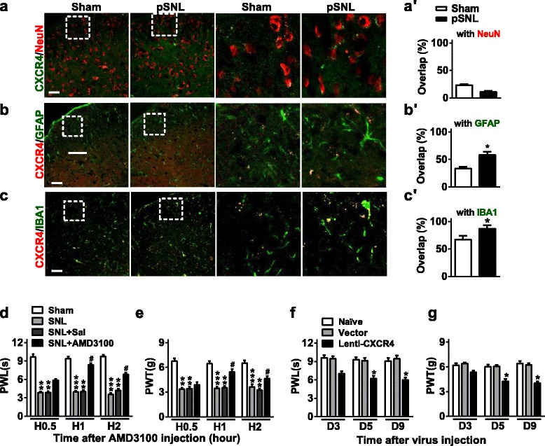
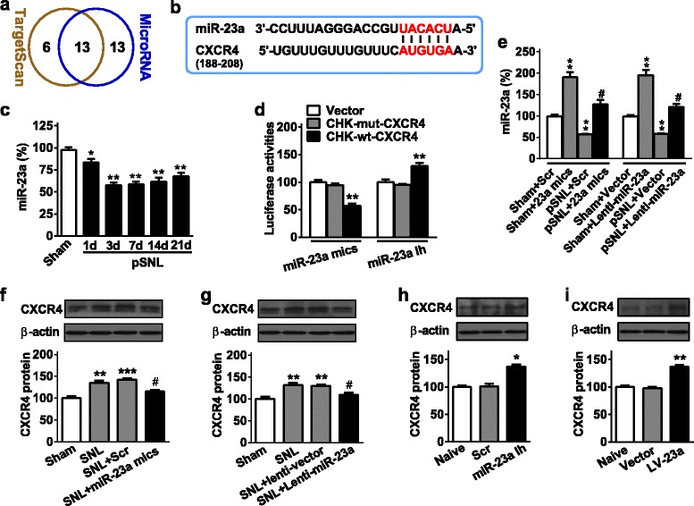
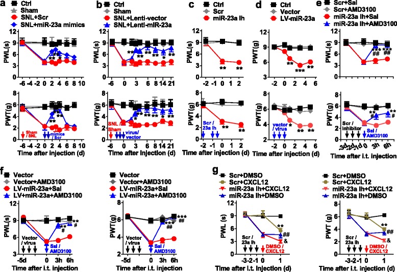
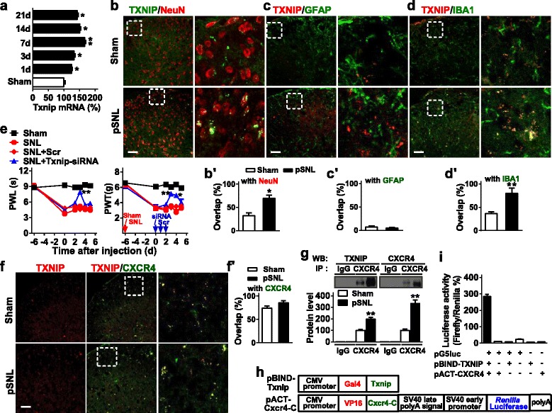
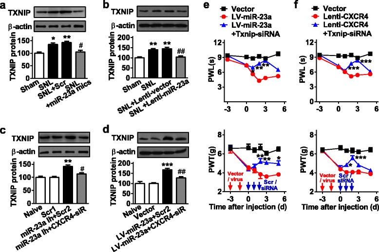
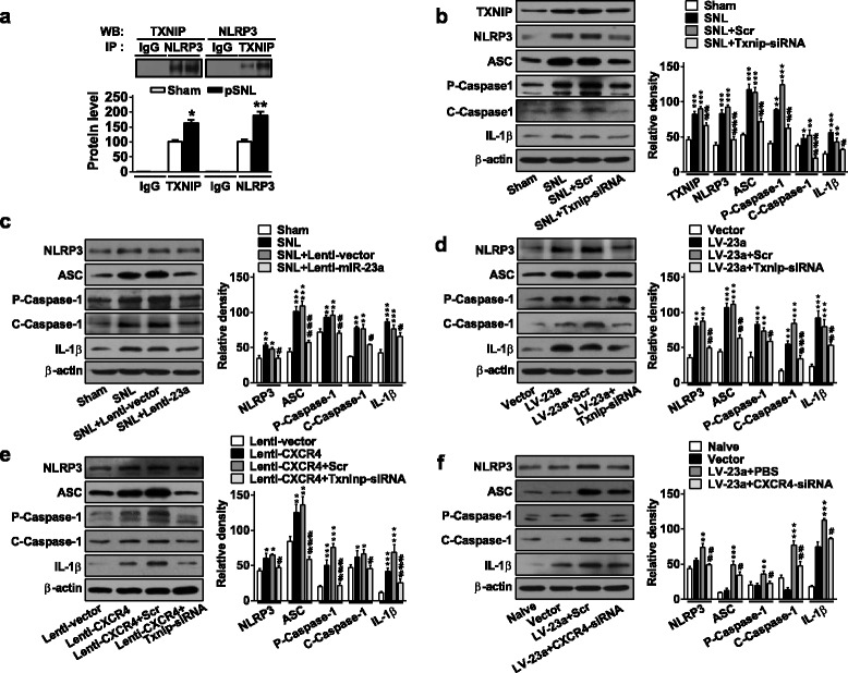
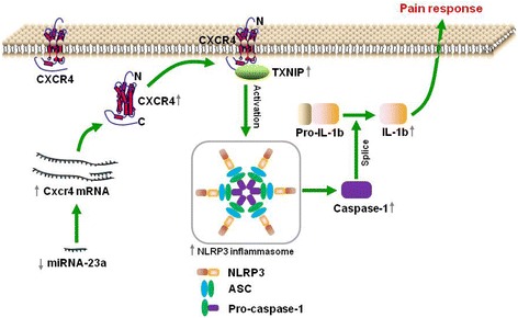
Results: CXCR4 expression increased in spinal glial cells of mice with pSNL-induced neuropathic pain. Blocking CXCR4 alleviated the pain behavior; contrarily, overexpressing CXCR4 induced pain hypersensitivity. MicroRNA-23a-3p (miR-23a) directly bounds to 3' UTR of CXCR4 mRNA. pSNL-induced neuropathic pain significantly reduced mRNA expression of miR-23a. Overexpression of miR-23a by intrathecal injection of miR-23a mimics or lentivirus reduced spinal CXCR4 and prevented pSNL-induced neuropathic pain. In contrast, knockdown of miR-23a by intrathecal injection of miR-23a inhibitor or lentivirus induced pain-like behavior, which was reduced by CXCR4 inhibition. Additionally, miR-23a knockdown or CXCR4 overexpression in naïve mice could increase the thioredoxin-interacting protein (TXNIP), which was associated with induction of NOD-like receptor protein 3 (NLRP3) inflammasome. Indeed, CXCR4 and TXNIP were co-expressed. The mammal two-hybrid assay revealed the direct interaction between CXCR4 and TXNIP, which was increased in the spinal cord of pSNL mice. In particular, inhibition of TXNIP reversed pain behavior elicited by pSNL, miR-23a knockdown, or CXCR4 overexpression. Moreover, miR-23a overexpression or CXCR4 knockdown inhibited the increase of TXNIP and NLRP3 inflammasome in pSNL mice.

Conclusions: miR-23a, by directly targeting CXCR4, regulates neuropathic pain via TXNIP/NLRP3 inflammasome axis in spinal glial cells. Epigenetic interventions against miR-23a, CXCR4, or TXNIP may potentially serve as novel therapeutic avenues in treating peripheral nerve injury-induced nociceptive hypersensitivity.

Keywords: CXCR4; NLRP3 inflammasome; Sciatic nerve injury; Spinal glia cell; TXNIP; miRNA-23a.


Conflict of interest statement

Ethics approval

All procedures were approved by the Committee on the Use of Live Animals in Teaching and Research and performed according to the guidelines for the care and use of laboratory animals as established by the Laboratory Animal Unit at the University of Hong Kong.

Consent for publication

Not applicable.

Competing interests

The authors declare that they have no competing interests.

Publisher’s Note

Springer Nature remains neutral with regard to jurisdictional claims in published maps and institutional affiliations.

Figures

Fig. 1
Fig. 1
CXCR4 modulates neuropathic pain. ac CXCR4 immunofluorescent co-staining with NeuN (a neuron marker) (a, a′), GFAP (an astrocyte marker) (b, b′) or IBA1 (a microglial marker) (c, c′) in the lumbar segment of the spinal cord at 7 days after pSNL surgery. *p < 0.05, versus Sham group by two-tailed unpaired Student’s t test; n = 5 per group. Original magnification × 200 for all the confocal images. Scale bar, 50 μm. d, e The effect of intrathecal injection of CXCR4 antagonist AMD3100 on the pSNL-induced thermal hyperalgesia (PWL) (d) and mechanical allodynia (PWT) (e) on day 7 after pSNL. Behavior was tested at 0.5, 1, and 2 h after AMD3100 injection. One-way ANOVA (behavior change versus treatment groups) followed by post hoc Tukey test. PWL F (6, 48) = 5.624; PWT F (6, 48) = 4.071, **p < 0.01, ***p < 0.001 versus Sham group; #p < 0.05 versus pSNL+Sal group. f, g Daily intrathecal injections of Lenti-CXCR4 for 3 consecutive days in naïve mice induced thermal hyperalgesia (f) and mechanical allodynia (g). Behavior was tested 48 h after lentivirus injection in naïve mice. One-way ANOVA (behavior change versus treatment groups) followed by post hoc Tukey test. PWL F (4, 36) = 57.264; PWT F (4, 36) = 3.194, *p < 0.05, versus vector group. Data are presented as mean ± SEM; n = 5 per group
Fig. 2
Fig. 2
miR-23a regulates CXCR4 expression in spinal astrocyte in neuropathic pain. a The shared miRNA numbers to targeting CXCR4 predicted by the use of TargetScan and MicroRNA program. b The informatics analysis of miR-23a binding the 3′UTR in CXCR4 mRNA. c Time course of spinal miR-23a expression in pSNL-induced chronic neuropathic pain mice. One-way ANOVA (expression versus time point) followed by post hoc Tukey test, Ftime (5, 24) = 19.66, *p < 0.05, **p < 0.01 versus sham group. d In vitro validation of miR-23a targeting CXCR4. A mutation was generated in the CXCR4-3′-UTR mRNA sequence in the complementary site for the seed region of miR-23a as indicated (CHK-mut-CXCR4). **p < 0.01 versus the corresponding CHK-mut-CXCR4 or empty vector group by two-tailed unpaired Student’s t test. e The validation of transfection efficiency of miR-23 mimics or Lenti-miR-23a in the mouse spinal cord by qRT-PCR. Spinal cord was harvested 24 h after intrathecal injection of continuous 2-day miR-23a mimics in naïve mice or pSNL mice with 7-day surgery or 72 h after intrathecal injection of continuous 2-day Lenti-miR-23a in naïve mice or pSNL mice with 7-day surgery. f, g The increased spinal CXCR4 protein expression in pSNL mice was reversed by intrathecal injection of miR-23a mimics (f) or Lenti-miR-23a (g). Intrathecal injections of miR-23a mimics or Lenti-miR-23a were performed from day 7 after pSNL. CXCR4 was measured at 24 h after 2-day miR-23a mimics injections or 48 h after 3-day Lenti-miR-23a injections. One-way ANOVA (expression versus treatment groups) followed by post hoc Tukey test, f F (3, 16) = 11.2, g Ftime (3, 16) = 14.6, **p < 0.01, ***p < 0.001, sham group; #p < 0.05 versus pSNL+Scr or pSNL+Lenti-vector group. h, i Spinal CXCR4 protein expression was increased by intrathecal injection of miR-23a inhibitor (miR-23a Ih) (h) or LV-miR-23a (LV-23a) (i) in naïve mice. CXCR4 was measured at 24 h after 2-day miR-23a inhibitor injections or at 48 h after 3-day LV-miR-23a injections. One-way ANOVA (expression versus treatment groups) followed by post hoc Tukey test, h G (2, 12) = 26.98, i H (2, 12) = 24.56, *p < 0.05, **p < 0.01 versus Scr or vector group. Data are presented as mean ± SEM; n = 5 per group
Fig. 3
Fig. 3
Spinal miR-23a regulates pain behavior via CXCR4. a, b Daily intrathecal injections of miR-23a mimics (a) or Lenti-miR-23a (b) for 2 or 3 consecutive days, respectively, reversed pSNL-induced thermal hyperalgesia and mechanical allodynia. Two-way ANOVA (effect versus group × time interaction) followed by post hoc Tukey test, a PWL Fgroup (18, 112) = 10.24, PWT Fgroup (18, 112) = 12.02; b PWL Fgroup (18, 112) = 10.57, PWT Fgroup (18, 112) = 8.12, *p < 0.05, **p < 0.01 versus pSNL+Scr or pSNL+Lenti-vector group. c, d Daily intrathecal injections of c miR-23a inhibitor or d LV-miR-23a for 2.5 or 3 consecutive days, respectively, produced thermal hyperalgesia and mechanical allodynia in naïve mice. Two-way ANOVA (effect versus group × time interaction) followed by post hoc Tukey test, c PWL Fgroup (4, 36) = 26.64, PWT Fgroup (4, 36) = 24.42; d PWL Fgroup (8, 60) = 4.83, PWT Fgroup (8, 60) = 5.31, **p < 0.01, ***p < 0.001 versus vector group. e, f Blocking CXCR4 with AMD3100 significantly reversed thermal hyperalgesia and mechanical allodynia induced by miR-23a inhibitor (e) or LV-miR-23a (f) in naïve mice. Two-way ANOVA (effect versus group × time interaction) followed by post hoc Tukey test, e PWL Fgroup (9, 64) = 27.84, PWT Fgroup (9, 64) = 12.69; f PWL Fgroup (9, 64) = 11.10, PWT Fgroup (9, 64) = 14.55, **p < 0.01, ***p < 0.001 versus 0 h before AMD3100 injection. #p < 0.05, ##p < 0.01 versus miR-23aIh + Sal or Lv-23a + Sal. Red arrow indicates pSNL surgery day. Blue or black arrow indicates injection time points of specified drug. Data are presented as mean ± SEM; n = 5 per group. g Intrathecal injection of CXCL12 further increased the thermal and mechanical sensitivity induced by miR-23a knockdown, with miR-23a inhibitor, in naïve mice. PWL Fgroup (6, 48) = 31.74, PWT Fgroup (6, 48) = 21.87; **p < 0.01 versus Scr + DMSO. ##p < 0.01 versus Scr + DMSO. &p < 0.05 versus miR-23a Ih + CXCL12. Black arrow indicates miR-23 inhibitor or Src injection. Red arrow indicates injection time points of CXCL12 or control DMSO. Data are presented as mean ± SEM; n = 5 per group
Fig. 4
Fig. 4
TXNIP is involved in the process of neuropathic pain by the CXCR4-dependent regulation. a Quantitative expression of Txnip mRNA at 1, 3, 7, 14, and 21 days after pSNL surgery. One-way ANOVA (expression versus time point) followed by post hoc Tukey test, Ftime (5, 24) = 40.4, *p < 0.05, **p < 0.01 versus sham group; n = 5 per group. bd TXNIP immunofluorescent co-staining with NeuN (b and b′), GFAP (c and c′), or IBA1 (d and d′) in the lumbar of spinal cord at 7 days after pSNL surgery. *p < 0.05, **p < 0.01 versus sham group by two-tailed unpaired Student’s t test; n = 5 per group. Scale bar, 50 μm. e Daily intrathecal injections of TXNIP siRNA for 3 consecutive days reversed pSNL-induced thermal hyperalgesia and mechanical allodynia. Two-way ANOVA (effect versus group × time interaction) followed by post hoc Tukey test, PWL Fgroup (10, 72) = 20.76; PWT Fgroup (10, 72) = 6.85, *p < 0.05, **p < 0.001, versus pSNL+Scr group; n = 5 per group. f, f′ Co-staining of double immunofluorescence (CXCR4, green; TXNIP, red) in the lumbar segment of the spinal cord of naïve mice. Scale bar, 50 μm. g The representative immunoblot demonstrates the interaction of TXNIP and CXCR4 in a co-IP experiment. Rabbit IgG is used as control for co-IP assays. **p < 0.01, ***p < 0.001 versus the corresponding sham group by two-tailed unpaired Student’s t test; n = 5 per group. h The CDS region of gene Txnip was inserted into pBIND to produce Gal4-Txnip chimeric fusion expression vector (pBIND-Txnip), and the C-terminal of Cxcr4 was inserted into pACT to produce VP16-Cxcr4-C chimeric fusion expression vector (pACT-CXCR-C). i pBIND-Txnip and/or pACT-Cxcr4-C transfected into 293T cells with the reporter gene vector pG5luc or empty vector. At 48 h after transfection, the interactions were measured by relative luciferase activity. In general, only pBIND-Txnip and pACT-Cxcr4-C co-transfection with pG5luc indicated strong luciferase activity. One-way ANOVA (expression versus the treated groups) followed by post hoc Tukey test, F (5, 24) = 628, ***p < 0.001; n = 3 per group
Fig. 5
Fig. 5
miR-23a/CXCR4 regulates neuropathic pain via TXNIP. a, b Increased spinal TXNIP protein expression in pSNL mice was reversed by intrathecal injection of miR-23a mimics (a) or Lenti-miR-23a (b). Intrathecal injections began on day 7 after pSNL. TXNIP was measured at 24 h after 2-day miR-23a mimics injections or at 48 h after 3-day Lenti-miR-23a injections. One-way ANOVA (expression versus treatment groups) followed by post hoc Tukey test a F (3, 16) = 12.54; b F (3, 16) = 17.27, *p < 0.01, **p < 0.01 versus sham group. #p < 0.05, ##p < 0.01 versus pSNL+Scr or pSNL+Lenti-vector group. n = 5 per group. c, d TXNIP protein expression increased by intrathecal injections of miR-23a Ih (c) or LV-miR-23a (d) in naïve mice was inhibited by intrathecal injection of CXCR4 pool siRNA. Intrathecal injection of CXCR4 pool siRNA was performed at 24 h after miR-23a Ih injections for 2 consecutive days or 48 h after LV-miR-23a injections for 3 consecutive days. TXNIP was measured at 24 h after CXCR4 pool siRNA injection. One-way ANOVA (expression versus treatment groups) followed by post hoc Tukey test c F (3, 16) = 18.1; d F (3, 16) = 31.69, **p < 0.01, ***p < 0.001 versus vector group. ##p < 0.01 versus miR-23a Ih or LV-miR-23a group. n = 5 per group. e Daily intrathecal injections of TXNIP siRNA for 3 consecutive days reversed pain-like behavior induced by LV-miR-23a. Two-way ANOVA (effect versus group × time interaction) followed by post hoc Tukey test, PWL Fgroup (10, 72) = 13.05; PWT Fgroup (10, 72) = 3.68. *p < 0.05, **p < 0.001 versus Lenti-miR-23a group; n = 5 per group. f Daily intrathecal injections of TXNIP siRNA for 3 consecutive days reversed pain-like behavior induced by Lenti-CXCR4. Two-way ANOVA (effect versus group × time interaction) followed by post hoc Tukey test, PWL Fgroup (10, 72) = 20.76; PWT Fgroup (10, 72) = 6.85, *p < 0.05, ***p < 0.0001 versus Lenti-CXCR4 group; n = 5 per group. Red arrow indicates pSNL day or vector/virus injection time points. Blue arrow indicates injection time points of Scr/siRNA. Data are presented as mean ± SEM
Fig. 6
Fig. 6
TXNIP regulates the protein expression of spinal NLRP3 inflammasome in SNL-induced neuropathic pain. a Co-IP of TXNIP and NLRP3. Rabbit IgG is used as control for co-IP assays. *p < 0.05, **p < 0.01 versus the corresponding sham group by two-tailed unpaired Student’s t test; n = 5 per group. b Increased NLRP3 inflammasome complex after pSNL, including NLRP3, ASC, P-Caspase1, C-Caspase1, and mature IL-1β, were reversed by knockdown of TXNIP with siRNA. Inflammasome complex expression was determined on day 7 after pSNL or at 24 h after 3-day injections of TXNIP siRNA or Scr, beginning on day 7 after pSNL. One-way ANOVA (expression versus the treated groups) followed by post hoc Tukey test, F (3, 8) = 76.56 for TXNIP, 106.9 for NLRP3, 79.01 for ASC, 191.3 for P-Caspase1, 26.68 for C-Caspase1, and 33.34 for mature IL-1β, **p < 0.01, ***p < 0.001 versus sham group. #p < 0.05, ##p < 0.01, and ###p < 0.001 versus pSNL+Scr group. c Increased NLRP3 inflammasome were reversed by intrathecal injection of Lenti-miR-23a in pSNL mice. Inflammasome complex expressions were measured at 7 days after pSNL surgery or at 48 h after 3-day injections of Lenti-miR-23a or Lenti-vector, beginning on day 7 after pSNL. One-way ANOVA (expression versus the treated groups) followed by post hoc Tukey test, F (3, 8) = 20.79 for NLRP3, 96.78 for ASC, 29.35 for P-Caspase1, 106.1 for C-Caspase1, and 42.21 for mature IL-1β, **p < 0.01, ***p < 0.001 versus sham group. #p < 0.05, ##p < 0.01, and ###p < 0.001 versus pSNL+Lenti-miR-23a group. d LV-miR-23a-induced expression of NLRP3 inflammasome was reversed by knockdown of TXNIP with siRNA in naïve mice. Content of inflammasome was examined at 48 h after 3-day injections of LV-miR-23a or at 24 h after 3-day injections of TXNIP siRNA or Scr (starting after the injections LV-miR-23a or vector). One-way ANOVA (expression versus the treated groups) followed by post hoc Tukey test, F (3, 8) = 106.7 for NLRP3, 138.4 for ASC, 54.04 for P-Caspase1, 133 for C-Caspase1, and 50.59 for mature IL-1β, **p < 0.01, ***p < 0.001 versus vector group. #p < 0.05, ##p < 0.01, and ###p < 0.001 versus LV-miR-23a + Scr group. e Increased NLRP3 inflammasome induced by injection of Lenti-CXCR4 was reversed by injection of TXNIP siRNA in naïve mice. Inflammasome complex expressions were measured at 48 h after 3-day injections of Lenti-CXCR4 or at 24 h after 3-day injection of TXNIP siRNA or Scr (starting after injections of Lenti-CXCR4 or Lenti-vector). One-way ANOVA (expression versus the treated groups) followed by post hoc Tukey test, F (3, 8) = 18.84 for NLRP3, 48.48 for ASC, 84.25 for P-Caspase1, 8.63 for C-Caspase1, and 48.37 for mature IL-1β, **p < 0.01, ***p < 0.001 versus Lenti-vector group. #p < 0.05, ##p < 0.01, and ###p < 0.001 versus Lenti-CXCR4 + Scr group. f NLRP3 inflammasome upregulated by injection of LV-miR-23a was reversed by injection of CXCR4 siRNA in naïve mice. One-way ANOVA (expression versus the treated groups) followed by post hoc Tukey test, F (3, 8) = 42.41 for NLRP3, 72.1 for ASC, 10.9 for P-Caspase1, 82.57 for C-Caspase1, and 290.7 for mature IL-1β, **p < 0.01, ***p < 0.001 versus vector group. #p < 0.05, ##p < 0.01 versus LV-miR-23a + Scr group. Expression of inflammasome was measured at 48 h after 3-day injections of LV-miR-23a or at 6 h after injection of CXCR4 siRNA or Scr (beginning after injections of LV-miR-23a or Vector). Data are presented as mean ± SEM; n = 3 per group
Fig. 7
Fig. 7
The schematic of miR-23a targeting CXCR4 regulates neuropathic pain via TXNIP/NLRP3 inflammasome in spinal glial cells of mice. In pSNL-induced chronic neuropathic pain, spinal miR-23a expression was significantly reduced, which increased the expression of spinal CXCR4, and subsequently the expression of TXNIP and NLRP3 inflammasome including NLRP3, ASC, Caspase-1, and IL-1β

Similar articles

Cited by

References

    1. Li M, Ransohoff RM. Multiple roles of chemokine CXCL12 in the central nervous system: a migration from immunology to neurobiology. Prog Neurobiol. 2008;84:116–131. doi: 10.1016/j.pneurobio.2007.11.003. - DOI - PMC - PubMed
    1. Bhangoo SK, Ren D, Miller RJ, Chan DM, Ripsch MS, Weiss C, McGinnis C, White FA. CXCR4 chemokine receptor signaling mediates pain hypersensitivity in association with antiretroviral toxic neuropathy. Brain Behav Immun. 2007;21:581–591. doi: 10.1016/j.bbi.2006.12.003. - DOI - PMC - PubMed
    1. Dubovy P, Klusakova I, Svizenska I, Brazda V. Spatio-temporal changes of SDF1 and its CXCR4 receptor in the dorsal root ganglia following unilateral sciatic nerve injury as a model of neuropathic pain. Histochem Cell Biol. 2010;133:323–337. doi: 10.1007/s00418-010-0675-0. - DOI - PubMed
    1. Knerlich-Lukoschus F, von der Ropp-Brenner B, Lucius R, Mehdorn HM, Held-Feindt J. Spatiotemporal CCR1, CCL3(MIP-1alpha), CXCR4, CXCL12(SDF-1alpha) expression patterns in a rat spinal cord injury model of posttraumatic neuropathic pain. J Neurosurg Spine. 2011;14:583–597. doi: 10.3171/2010.12.SPINE10480. - DOI - PubMed
    1. Luo X, Tai WL, Sun L, Pan Z, Xia Z, Chung SK, Cheung CW. Crosstalk between astrocytic CXCL12 and microglial CXCR4 contributes to the development of neuropathic pain. Mol Pain. 2016;12 - PMC - PubMed

MeSH terms

LinkOut - more resources