. 2018 Jan 31;17(1):18.
doi: 10.1186/s12943-018-0761-9.

Long non-coding RNA XLOC_000647 suppresses progression of pancreatic cancer and decreases epithelial-mesenchymal transition-induced cell invasion by down-regulating NLRP3

Affiliations

Long non-coding RNA XLOC_000647 suppresses progression of pancreatic cancer and decreases epithelial-mesenchymal transition-induced cell invasion by down-regulating NLRP3

Hao Hu et al. Mol Cancer. .

Abstract

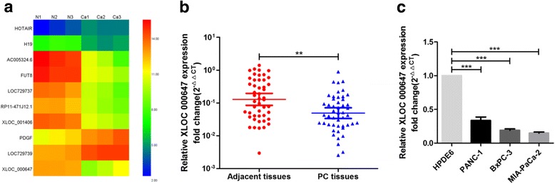
Background: Long non-coding RNAs (lncRNAs) play an important role in the development and progression of various tumors, including pancreatic cancer (PC). Recent studies have shown that lncRNAs can 'act in cis' to regulate the expression of its neighboring genes. Previously, we used lncRNAs microarray to identify a novel lncRNA termed XLOC_000647 that was down-regulated in PC tissues. However, the expression and function of XLOC_000647 in PC remain unclear.

Methods: The expression of XLOC_000647 and NLRP3 in PC specimens and cell lines were detected by quantitative real-time PCR. Transwell assays were used to determine migration and invasion of PC cells. Western blot was carried out for detection of epithelial-mesenchymal transition (EMT) markers in PC cells. The effect of XLOC_000647 on PC cells was assessed in vitro and in vivo. The function of NOD-like receptor family pyrin domain-containing 3 (NLRP3) in PC was investigated in vitro. In addition, the regulation of NLRP3 by XLOC_000647 in PC was examined in vitro.

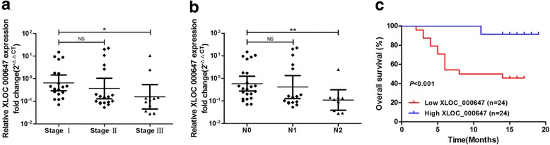
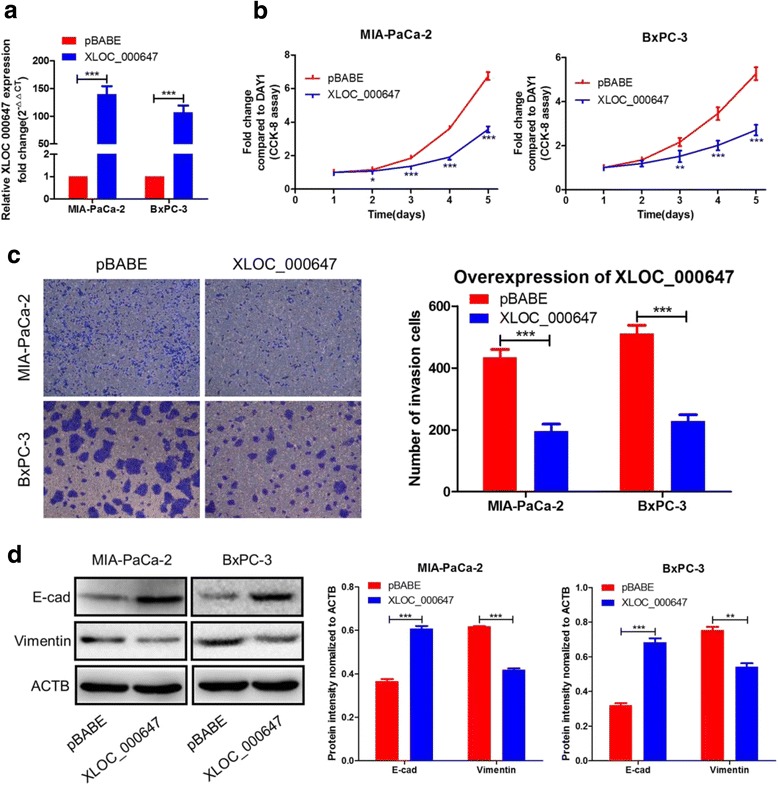
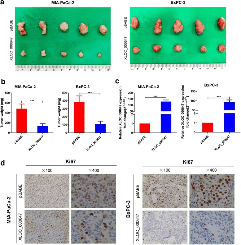
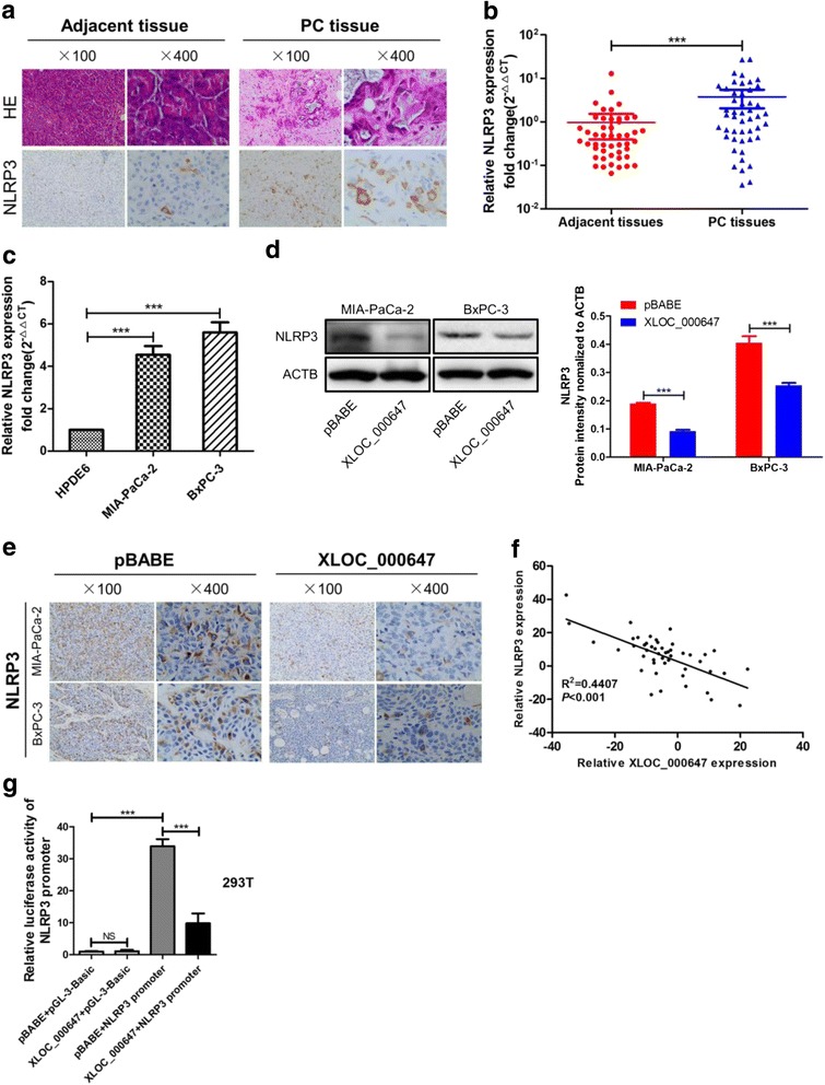
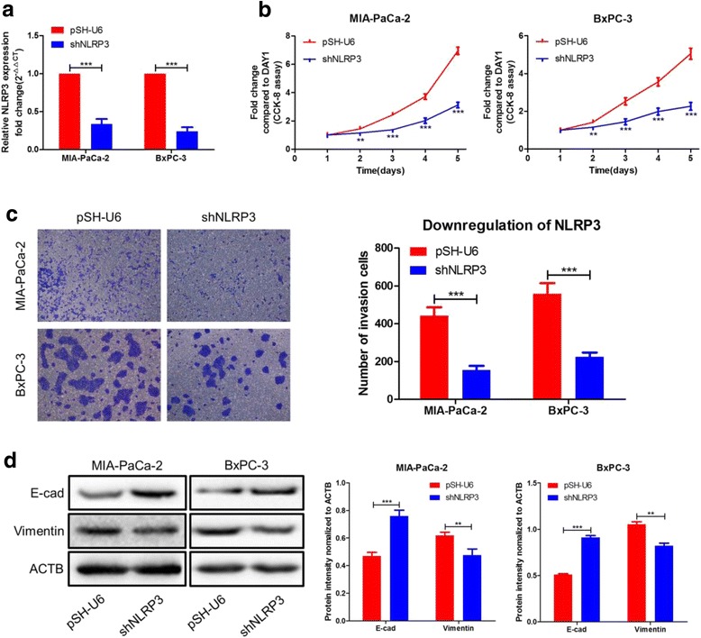
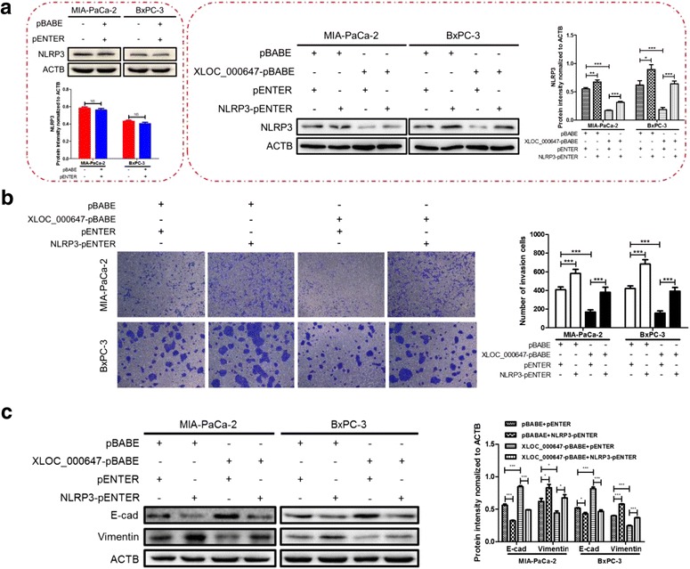
Results: Here, XLOC_000647 expression was down-regulated in PC tissues and cell lines. The expression level of XLOC_000647 was significantly correlated to tumor stage, lymph node metastasis, and overall survival. Overexpression of XLOC_000647 attenuated cell proliferation, invasion, and EMT in vitro and impaired tumor growth in vivo. Further, a significantly negative correlation was observed between XLOC_000647 levels and its genomic nearby gene NLRP3 in vitro and in vivo. Moreover, XLOC_000647 decreased NLRP3 by inhibiting its promoter activity. Knockdown of NLRP3 decreased proliferation of cancer cells, invasion, and EMT in vitro. Importantly, after XLOC_000647 was overexpressed, the corresponding phenotypes of cells invasion and EMT were reversed by overexpression of NLRP3.

Conclusions: Together, these results indicate that XLOC_000647 functions as a novel tumor suppressor of lncRNA and acts as an important regulator of NLRP3, inhibiting cell proliferation, invasion, and EMT in PC.

Keywords: Epithelial mesenchymal transition; Invasion; LncRNAs; NLRP3; Pancreatic cancer.


Conflict of interest statement

Ethics approval and consent to participate

The clinical protocol was approved by the Ethics Committee of The First Hospital Affiliated to Nanjing Medical University and all patients signed a written informed consent form before specimen collection. The animal care and experimental protocols were approved by the institutional guidelines of Jiangsu Province and by the Animal Care and Use Committee of Nanjing Medical University.

Consent for publication

Not applicable.

Competing interests

The authors declare that they have no competing interests.

Publisher’s Note

Springer Nature remains neutral with regard to jurisdictional claims in published maps and institutional affiliations.

Figures

Fig. 1
Fig. 1
Expression of XLOC_000647 in pancreatic cancer (PC) tissues and cell lines. a Heat maps from our previous lncRNAs tissues microarray. N represents normal pancreatic tissue and Ca represents PC tissue. b Relative XLOC_000647 expression in tissues (n = 48). XLOC_000647 expression from all tissues was normalized to ACTB expression (ΔCT) and then compared with an adjacent tissue and converted to the fold change (2−ΔΔCT). c Relative XLOC_000647 expression in cell lines. Data are shown as fold change (2−ΔΔCT) and the mean ± SD from three independent experiments. **P < 0.01, ***P < 0.001
Fig. 2
Fig. 2
Associations between XLOC_000647, clinicopathologic characteristics and prognosis after surgery. a XLOC_000647 expression in different TNM stages of PC, stages I, II, and III (n = 19, n = 18 and n = 11, respectively). b XLOC_000647 expression in the lymph node metastasis- N0 group (N0, n = 23), -N1 group (N1, n = 16) and -N2 group (N2, n = 9). The qRT-PCR data are shown as fold change (2−ΔΔCT). For expressions in tissues, the levels were firstly normalized to ACTB expression as ΔCT and then compared with one of the tissues and converted to the fold change (2−ΔΔCT). c Overall survival curve of the high-level and low-level group divided by XLOC_000647 expression. The P-values are shown with the log-rank test (two-sided). NS (not significant). *P < 0.05, **P < 0.01, ***P < 0.001
Fig. 3
Fig. 3
Influence of XLOC_000647 on the PC cell proliferation, cell invasion, and EMT. a The expression of XLOC_000647 was up-regulated by XLOC_000647-pBABE in MIA-PaCa-2 and BxPC-3 cells. b The proliferation activity of the two cell lines detected by CCK-8 assay. c The invasion capacity of the two cell lines when compared with the controls by transwell assays. d Analysis of the E-cad and Vimentin protein levels in the two cell lines and corresponding control cells by western blot. Results are represented as protein intensity relative to ACTB. *P < 0.05, **P < 0.01, ***P < 0.001
Fig. 4
Fig. 4
Stable overexpression of XLOC_000647 significantly reduces tumor growth in vivo. a Tumors removed from the mice 6 weeks after injection of MIA-PaCa-2 or BxPC-3 cells stably transfected with XLOC_000647-pBABE or pBABE, respectively. b Tumor weights are shown as the means ± SD when the tumors were harvested. c The expression of XLOC_000647 in paired tumor tissues was analyzed by qRT-PCR. d Representative images (×100 and ×400) of IHC staining of the tumor. Results showed that overexpression of XLOC_000647 decreased the proliferation index of Ki67. ***P < 0.001
Fig. 5
Fig. 5
Expression of NLRP3 in PC tissues and cell lines, and association between NLRP3 and XLOC_000647. a Representative images (×100 and ×400) of H and E (hematoxylin and eosin) and immunohistochemical (IHC) staining for NLRP3 in paraffin-embedded PC and corresponding adjacent tissues. b Relative XLOC_00067 expression in tissues (n = 48). PC tissues versus corresponding adjacent tissues. c Relative XLOC_000647 expression in cell lines and the mean ± SD from three independent experiments. d Influence of XLOC_000647-stable overexpression on the expression level of NLRP3 in cell lines by western blot. e Representative images (×100 and ×400) of IHC staining of the tumor from mice. Results showed that overexpression of XLOC_000647 decreased the expression level of NLRP3. f The correlation between NLRP3 mRNA levels and XLOC_00067 levels in 48 PC tissues (R2 = 0.4407, P < 0.001). g The luciferase activity of NLRP3 promoter is decreased by XLOC_000647 in 293 T cells. NS (not significant). ***P < 0.001
Fig. 6
Fig. 6
Influence of NLRP3 on PC cell proliferation, cell invasion, and EMT. a The expression of NLRP3 was down-regulated by shNLRP3 in MIA-PaCa-2 and BxPC-3 cells. b The proliferation activity of these two cell lines detected by CCK-8 assay. c The invasion capacity of the two cell lines when compared with the controls by transwell assays. d Analysis of the E-cad and Vimentin protein levels in the two cell lines and corresponding control cells by western blot. **P < 0.01, ***P < 0.001
Fig. 7
Fig. 7
Inhibition of EMT-induced cells invasion regulated by XLOC_000647 overexpression was reversed by NLRP3 overexpression. a The vectors of pBABE + pENTER alone had no effect on the expression of NLRP3 (left). The protein levels of NLRP3 were up-regulated after transfected with NLRP3-pENTER in cells of XLOC_000647-stable overexpression compared with control cells (right). b The invasion of XLOC_000647-overexpressed cells transfected with NLRP3-pENTER when compared with the controls by transwell assays. Values represented the mean ± SD from three independent experiments. c Analysis of the E-cad and Vimentin protein levels in XLOC_000647-overexpressed cells transfected with NLRP3-pENTER and pENTER by western blot. NS (not significant). *P < 0.05, **P < 0.01, ***P < 0.001

Similar articles

Cited by

References

    1. Waddell N, Pajic M, Patch AM, Chang DK, Kassahn KS, Bailey P, Johns AL, Miller D, Nones K, Quek K, et al. Whole genomes redefine the mutational landscape of pancreatic cancer. Nature. 2015;518:495–501. doi: 10.1038/nature14169. - DOI - PMC - PubMed
    1. Hessmann E, Johnsen SA, Siveke JT, Ellenrieder V. Epigenetic treatment of pancreatic cancer: is there a therapeutic perspective on the horizon? Gut. 2017;66:168–179. doi: 10.1136/gutjnl-2016-312539. - DOI - PMC - PubMed
    1. Zhan HX, JW X, Wu D, Zhang TP, SY H. Pancreatic cancer stem cells: new insight into a stubborn disease. Cancer Lett. 2015;357:429–437. doi: 10.1016/j.canlet.2014.12.004. - DOI - PubMed
    1. Ansari D, Tingstedt B, Andersson B, Holmquist F, Sturesson C, Williamsson C, Sasor A, Borg D, Bauden M, Andersson R. Pancreatic cancer: yesterday, today and tomorrow. Future Oncol. 2016;12:1929–1946. doi: 10.2217/fon-2016-0010. - DOI - PubMed
    1. Rinn JL, Chang HY. Genome regulation by long noncoding RNAs. Annu Rev Biochem. 2012;81:145–166. doi: 10.1146/annurev-biochem-051410-092902. - DOI - PMC - PubMed

Publication types

MeSH terms

Substances