. 2018 Jan 31;9(1):21.
doi: 10.1186/s13287-018-0768-6.

Curcumin-mediated bone marrow mesenchymal stem cell sheets create a favorable immune microenvironment for adult full-thickness cutaneous wound healing

Affiliations

Curcumin-mediated bone marrow mesenchymal stem cell sheets create a favorable immune microenvironment for adult full-thickness cutaneous wound healing

Zhi Yang et al. Stem Cell Res Ther. .

Abstract

Background: Adult full-thickness cutaneous wound repair suffers from an imbalanced immune response, leading to nonfunctional reconstructed tissue and fibrosis. Although various treatments have been reported, the immune-mediated tissue regeneration driven by biomaterial offers an attractive regenerative strategy for damaged tissue repair.

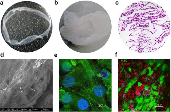
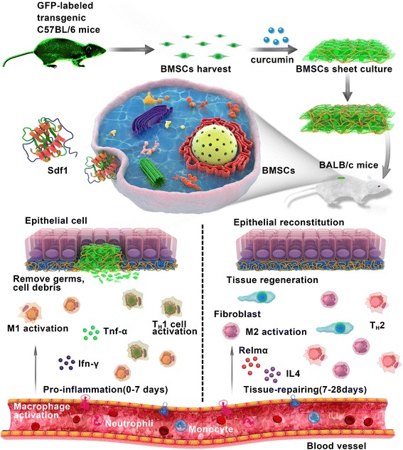
Methods: In this research, we investigated a specific bone marrow-derived mesenchymal stem cell (BMSC) sheet that was induced by the Traditional Chinese Medicine curcumin (CS-C) and its immunomodulatory effects on wound repair. Comparisons were made with the BMSC sheet induced without curcumin (CS-N) and control (saline).

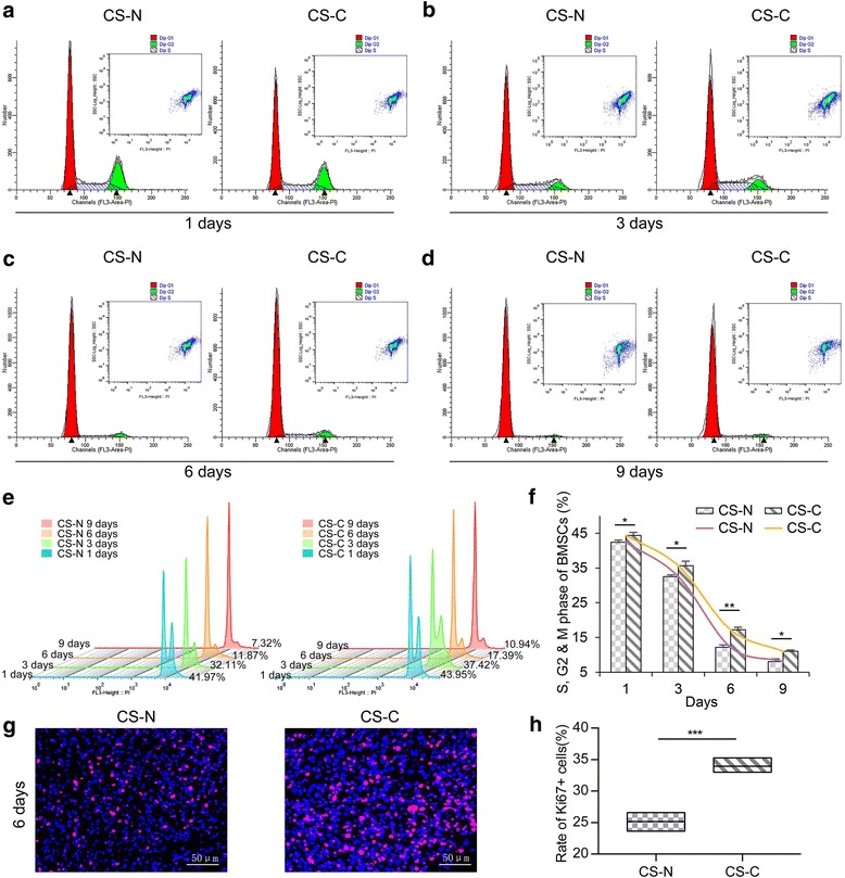
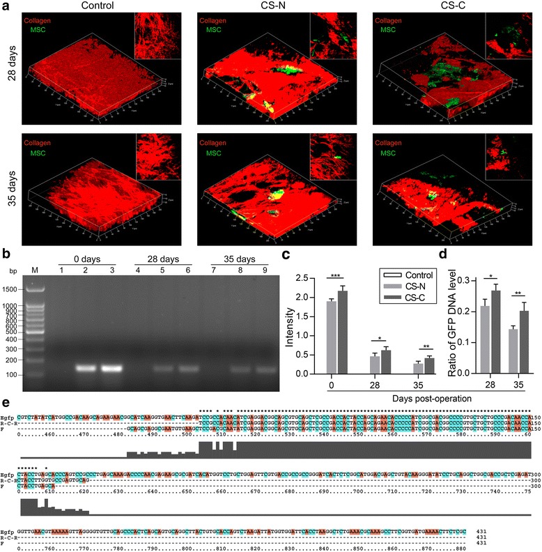
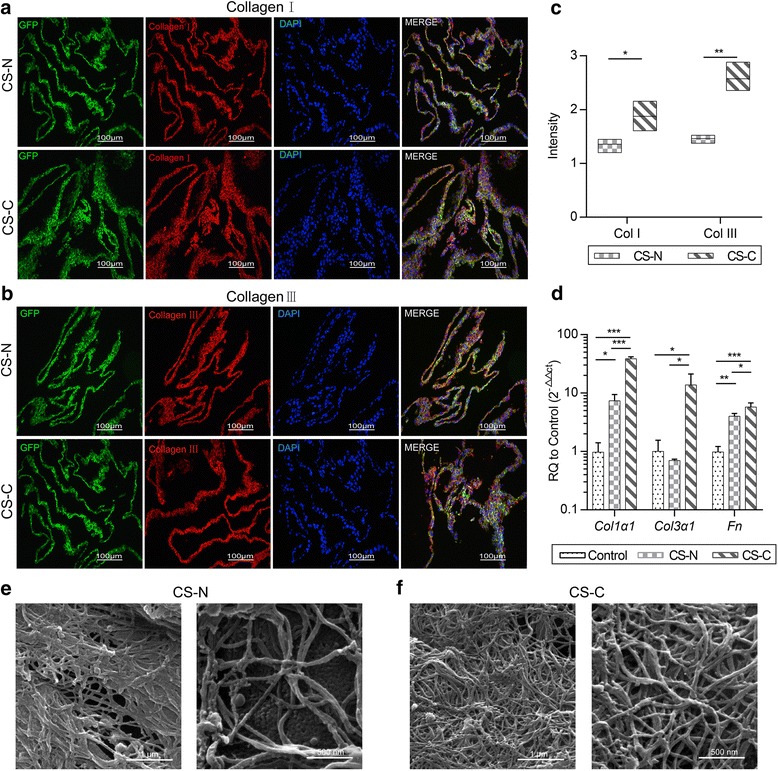
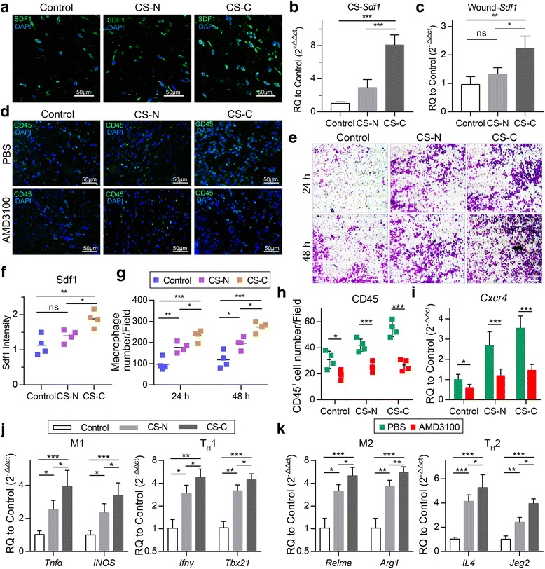
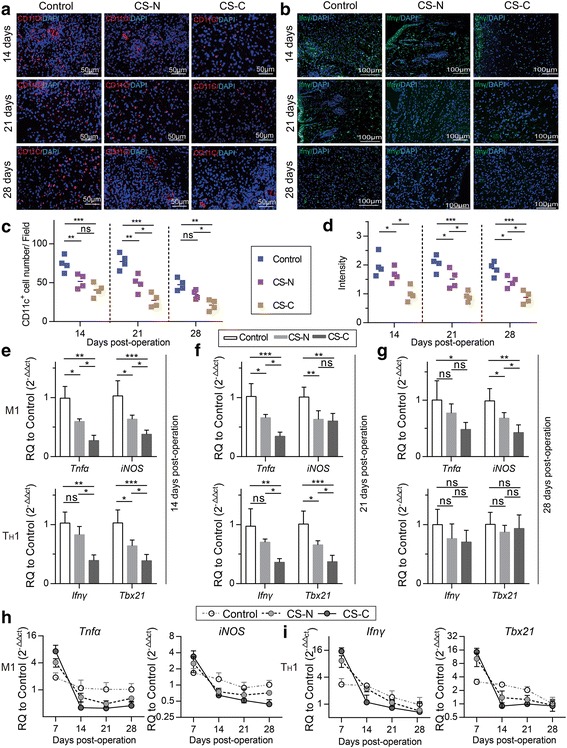
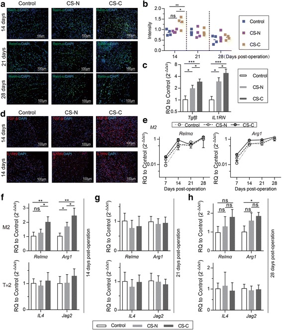
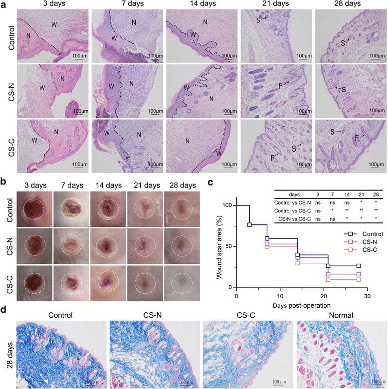
Results: In vitro cultured BMSC sheets (CS-C) showed that curcumin promoted the proliferation of BMSCs and modified the features of produced extracellular matrix (ECM) secreted by BMSCs, especially the contents of ECM structural proteins such as fibronectin (FN) and collagen I and III, as well as the ratio of collagen III/I. Two-photon fluorescence (TPF) and second-harmonic generation (SHG) imaging of mouse implantation revealed superior engraftment of BMSCs, maintained for 35 days in the CS-C group. Most importantly, CS-C created a favorable immune microenvironment. The chemokine stromal cell-derived factor 1 (SDF1) was abundantly produced by CS-C, thus facilitating a mass migration of leukocytes from which significantly increased expression of signature TH1 cells (interferon gamma) and M1 macrophages (tumor necrosis factor alpha) genes were confirmed at 7 days post-operation. The number of TH1 cells and associated pro-inflammatory M1 macrophages subsequently decreased sharply after 14 days post-operation, suggesting a rapid type I immune regression. Furthermore, the CS-C group showed an increased trend towards M2 macrophage polarization in the early phase. CS-C led to an epidermal thickness and collagen deposition that was closer to that of normal skin.

Conclusions: Curcumin has a good regulatory effect on BMSCs and this promising CS-C biomaterial creates a pro-regenerative immune microenvironment for cutaneous wound healing.

Keywords: Bone marrow mesenchymal stem cell; Cell sheet; Curcumin; Cutaneous wound healing; Immune response.


Conflict of interest statement

Ethics approval

All experimental protocols were approved by the Ethical Committee for Animal Experiments of South China Normal University. All animal experiments conducted in this research were performed in accordance with the guidelines of South China Normal University Intramural Animal Use and Care Committee and met the NIH guidelines for the care and use of laboratory animals.

Consent for publication

All authors have read and approved the manuscript for publication.

Competing interests

The authors declare that they have no competing interests.

Publisher’s Note

Springer Nature remains neutral with regard to jurisdictional claims in published maps and institutional affiliations.

Figures

Fig. 1
Fig. 1
Characterization of the BMSC sheet. a The appearance of the BMSC sheet (6 cm in diameter). b Stereomicroscope image of the BMSC sheet (5×). c H&E staining of BMSC sheets which contained many layers of cells. d Scanning electron microscope image of the BMSC sheet; the arrows point to mesenchymal stem cells (MSCs) (1 kx, 20 μm). e Fluorescence microscope image of the BMSC sheet; green and blue show the cytoskeleton of GFP+ BMSCs and cell nuclei, respectively (63×, 5 μm). f Second harmonic imaging (SHG) image of the BMSC sheet; red and green represent collagen and cells, respectively (40×, 20 μm)
Fig. 2
Fig. 2
Influence of curcumin on bone marrow-derived mesenchymal stem cell (BMSC) proliferation activity. BMSCs treated with curcumin at a concentration of 0.5 μM (CS-C) and without curcumin (CS-N) for 1 day (a), 3 days (b), 6 days (c), and 9 days (d), respectively. The cell cycle was determined by flow cytometry. e Flow cytometry to assess the cell cycle at the indicated intervals (n = 3). f Quantification of the number of MSCs at the S, G2, and M phase. g Immunofluorescence staining of the proliferation of cells with Ki67 in the CS-N and CS-C groups (cultured for 6 days). h Quantification of the rate of Ki67-positive cells in CS-N and CS-C groups (n = 4). Student’s t test: *P < 0.05, **P < 0.01, ***P < 0.001
Fig. 3
Fig. 3
Engraftment of GFP+ BMSCs during wound healing. a Second harmonic imaging (SHG) of control (left), BMSC sheets induced without curcumin (CS-N; middle), and BMSC sheets induced by curcumin (CS-C; right) transplanted into the wound site at 28 days and 35 days. Green and red indicate GFP+ mesenchymal stem cells (MSCs) and collagen, respectively. b The contents of GFP+ BMSCs represented by DNA agarose gel electrophoresis; each lane shows control, CS-N, and CS-C, respectively, at 0 days (lanes 1, 2, 3), 28 days (lanes 4, 5, 6), and 35 days (lanes 7, 8, 9). c Relative quantitative analysis of the content of GFP DNA (n = 4 mice/group). d Ratio of green fluorescent protein (GFP) DNA level at 28 days and 35 days to 0 days. e Sequence alignment results for the amplified products with GFP sequences. Student’s t test (d) and ANOVA (c): *P < 0.05, **P < 0.01, ***P < 0.001
Fig. 4
Fig. 4
Effect of curcumin on the BMSC sheets. a,b Immunohistochemical staining of the BMSC sheets induced without curcumin (CS-N) and BMSC sheets induced by curcumin (CS-C); green, red, and blue represent green fluorescent protein (GFP)+ cells, collagen I (a)/collagen III (b), and nuclei, respectively (20×, 100 μm) (n = 4). c Content variation in collagen (Col) I and collagen III in the CS-N group and CS-C group. d RT-PCR results for gene expression in the extracellular matrix, including col1α1, col3α1, and Fn. SEM image of the e CS-N group and f CS-C group (left is 20 kx, 1 μm, right is 40 kx, 500 nm). Student’s t test and ANOVA: *P < 0.05, **P < 0.01, ***P < 0.001
Fig. 5
Fig. 5
CS-C recruits macrophages and T cells in early repair stages dependent on SDF1. a Immunofluorescence staining of stromal cell-derived factor 1 (SDF1) in the wound area in the control, BMSC sheets induced without curcumin (CS-N), and BMSC sheets induced by curcumin (CS-C) groups at 7 days post-operation (40×, 50 μm) (n = 4 mice/group). b Gene expression of SDF1 in the control, CS-N, and CS-C groups in vitro. c Gene expression of SDF1 in the wound area at 7 days post-operation. d Immunofluorescence staining of CD45+ leukocytes in the wound area in the control, CS-N, and CS-C groups at 7 days post-operation with AMD3100 or without (phosphate-buffered saline (PBS) instead of AMD3100) (40×, 50 μm) (n = 4 mice/group). e Macrophage chemotaxis in the control, CS-N, and CS-C groups at 24 and 48 h (20×, 50 μm). f Quantification of the SDF1 intensity at 7 days-post-operation. g Quantification of the number of migrated macrophages. h Quantification of the number of CD45+ cells at 7 days post-operation. i Gene expression of Cxcr4 in the control, CS-N, and CS-C groups at 7 days post-operation with or without AMD3100 (n = 5 mice/group). j Expression levels of Tnfα, iNOS (signature genes of M1 macrophages), and Ifnγ, Tbx21 (signature genes of TH1 cells) by RT-PCR at 7 days-post-operation. k Expression levels of Relmα, Arg1 (signature genes of M2 macrophages), and IL4, Jag2 (signature genes of TH2cells) by RT-PCR (n = 5 mice/group). ANOVA: *P < 0.05, **P < 0.01, ***P < 0.001
Fig. 6
Fig. 6
CS-C accelerated type I immune reaction regression. a Immunofluorescence staining of CD11c+ M1 macrophages in the control, BMSC sheets induced without curcumin (CS-N), and BMSC sheets induced by curcumin (CS-C) groups at 14, 21, and 28 days after treatment. Red and blue represent CD11c+ M1 macrophages and nuclei, respectively (20×, 50 μm) (n = 4 mice/group). b Immunofluorescence staining of interferon (IFN)γ at 14, 21, and 28 days. Green and blue represent T helper 1 (TH1) cells and nuclei, respectively (bar = 100 μm) (n = 4 mice/group). Quantification of c the number of M1 macrophages and d TH1 cells, respectively. Expression levels of Tnfα and iNOS (signature genes of M1 macrophages) and Ifnγ and Tbx21 (signature genes of TH1 cells) by RT-PCR at 14 (e), 21 (f), and 28 days (g) after transplantation with saline, CS-N, and CS-C (n = 5 mice/group). M1 (h) and TH1 (i) signature gene expression during the whole experimental period for the control, CS-N, and CS-C groups. ANOVA: *P < 0.05, **P < 0.01, ***P < 0.001. ns not significant
Fig. 7
Fig. 7
CS-C accelerated M2 macrophage polarization. a Immunofluorescence staining of Relmα in the control, BMSC sheets induced without curcumin (CS-N), and BMSC sheets induced by curcumin (CS-C) groups at 14, 21, and 28 days after treatment. Red and blue represent Relmα and nuclei, respectively (20×, 100 μm) (n = 4 mice/group). b Quantification of the Relmα intensity. c Expression levels of Tgfβ and IL1RN by RT-PCR at 14 days post-operation (n = 5 mice/group). d Immunofluorescence staining of Tgfβ+ (top panels) and IL1RN+ (bottom panels) cells in the three groups at 14 days post-operation. Red represents Tgfβ and IL1RN, and blue represents nuclei (20×, 100 μm) (n = 4 mice/group). e M2 signature gene expression during the whole experimental period for the control, CS-N, and CS-C groups. Expression levels of Relmα and Arg1 (signature genes of M2 macrophages) and IL4 and Jag2 (signature genes of TH2 cells) by RT-PCR at 14 (f), 21 (g), and 28 days (h) after transplantation with saline, CS-N, and CS-C (n = 5 mice/group). ANOVA: *P < 0.05, **P < 0.01, ***P < 0.001. ns not significant
Fig. 8
Fig. 8
CS-C promotes skin reconstruction. a Immunohistochemical staining (10×, 100 μm) and b macroscopic observation of the wound areas in the control, BMSC sheets induced without curcumin (CS-N), and BMSC sheets induced by curcumin (CS-C) groups at days 3, 7, 14, 21, and 28 (n = 5 mice/group). c Statistical analysis of wound closure rate in continuous time (3, 7, 14, 21, and 28 days). d Masson’s trichrome staining (10×, 100 μm) of healing wounds in the control group, CS-N group, CS-C group, and normal group at 28 days post-operation. ANOVA: *P < 0.05, **P < 0.01, ***P < 0.001. F hair follicle, N normal site, ns not significant, S sebaceous gland, W wound site
Fig. 9
Fig. 9
The CS-C-based immunomodulatory process in wound healing. Green fluorescent protein (GFP)+ bone marrow-derived stem cells (BMSCs) were harvested from transgenic C57BL/6 mice. BMSC sheets were prepared by culturing 1.5 × 105 third passage cells on culture dishes for 12 days. The BMSC sheets were then transplanted into the skin wounds of the recipient mice. Once CS-C is applied to the wound site, it induces the secretion of various chemokines. Stromal cell-derived factor 1 (Sdf1) increases significantly, recruiting more leukocytes, such as macrophages (M) and T helper (TH) cells, to the wound area. Abundant type I immune cells of M1 macrophages and TH1 cells are activated at approximately 7 days of repair in the pro-inflammatory stage. The temporary pro-inflammatory response leads to the rapid removal of foreign pathogens. After 7 days post-operation, the typical type I immune response, the number of M1 macrophages and TH1 cells near the wound are greatly reduced; instead, there is an increase in type II immune response, as reflected in the number of anti-inflammatory M2 macrophages. Timely suppression of the type I immune reaction allows rapid tissue rebuilding, and the increase in type II anti-inflammatory M2 macrophages in the following stage is beneficial for tissue repairing. Through the transplantation of CS-C, rapid and effective skin wound healing occurs. Ifn interferon, IL interleukin, Tnf tumor necrosis factor

Similar articles

Cited by

References

    1. Stadelmann WK, Digenis AG, Tobin GR. Physiology and healing dynamics of chronic cutaneous wounds. Am J Surg. 1998;176:26S–38S. doi: 10.1016/S0002-9610(98)00183-4. - DOI - PubMed
    1. Falanga V. Wound healing and its impairment in the diabetic foot. Lancet. 2005;366:1736–1743. doi: 10.1016/S0140-6736(05)67700-8. - DOI - PubMed
    1. Wynn TA, Ramalingam TR. Mechanisms of fibrosis: therapeutic translation for fibrotic disease. Nat Med. 2012;18:1028–1040. doi: 10.1038/nm.2807. - DOI - PMC - PubMed
    1. Nagaraja S, Wallqvist A, Reifman J, Mitrophanov AY. Computational approach to characterize causative factors and molecular indicators of chronic wound inflammation. J Immunol. 2014;192:1824–1834. doi: 10.4049/jimmunol.1302481. - DOI - PubMed
    1. Duffield JS, Forbes SJ, Constandinou CM, Clay S, Partolina M, Vuthoori S, Wu S, Lang R, Iredale JP. Selective depletion of macrophages reveals distinct, opposing roles during liver injury and repair. J Clin Invest. 2005;115:56–65. doi: 10.1172/JCI200522675. - DOI - PMC - PubMed

Publication types

MeSH terms