. 2018 Jan 31;9(1):20.
doi: 10.1186/s13287-017-0745-5.

Effects of hPMSCs on granulosa cell apoptosis and AMH expression and their role in the restoration of ovary function in premature ovarian failure mice

Affiliations

Effects of hPMSCs on granulosa cell apoptosis and AMH expression and their role in the restoration of ovary function in premature ovarian failure mice

Hongqin Zhang et al. Stem Cell Res Ther. .

Retraction in

Abstract

Background: This study was performed to determine the effects of human placenta mesenchymal stem cell (hPMSC) transplantation on granulosa cell apoptosis and anti-Müllerian hormone (AMH) and follicle-stimulating hormone receptor (FSHR) expression in autoimmune drug-induced premature ovarian failure (POF) mice. The aim of this research is to investigate the mechanisms of hPMSCs on ovarian reserve capacity.

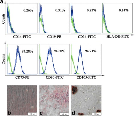
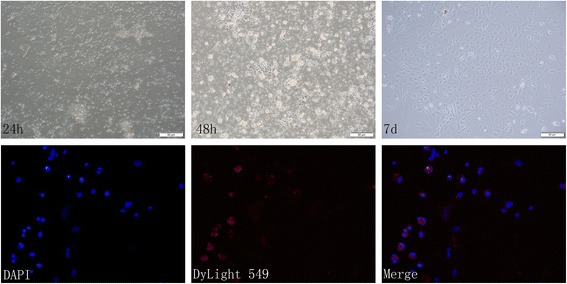
Methods: The POF mice model was established by injection of zona pellucida 3 peptide (pZP3). hPMSC transplantation was conducted by intravenous injection into mice following pZP3 treatment. The follicle number was examined by histopathology. The serum levels of FSH, LH, E2, AMH and anti-zona pellucida antibody (AzpAb) were measured by enzyme-linked immunosorbent assay. AMH and FSHR expression in the ovary was analyzed by immunohistochemistry and western blot analysis. Granulosa cell apoptosis of the ovaries was examined by In Situ Cell Death Detection Kit. Granulosa cells were isolated and treated with SiAmh interference and hPMSC supernatant to observe the effects of AMH expression on granulosa cell apoptosis in vitro.

Results: The results showed that hPMSC transplantation can significantly recover the estrus cycle in the POF group. Morphological staining showed that the basal follicles and sinus follicles after hPMSC transplantation were higher in POF mice than in those without treatment, and the follicle number was significantly decreased with atresia. The serum levels of FSH, LH and AzpAb in the hPMSC transplantation group were reduced considerably, but the E2 and AMH levels were significantly increased. After hPMSC transplantation, the AMH and FSHR expression in ovarian tissue was significantly higher than in the POF group as determined by immunochemistry and western blot analysis. The FSHR expression was shown in granulosa cells only, and FSHR expression increases with AMH expressed in the ovary; granulosa cell apoptosis was decreased following hPMSC transplantation. The same results were observed from the in-vitro study.

Conclusions: hPMSC transplantation can significantly improve the serum levels of high gonadotropin and low estrogen of POF mice, promote follicular development, inhibit excessive follicular atresia and granulosa cell apoptosis, and improve the ovarian reserve capacity. The mechanism may be achieved by increasing the expression of AMH and FSHR in ovaries.

Keywords: Anti-Müllerian hormone; Apoptosis; Follicle stimulating hormone receptor; Granulosa cells; Mesenchymal stem cell; Placenta; Premature ovarian failure.


Conflict of interest statement

Ethics approval

All animal care and experimental procedures were performed according to the guide for the Care and Use of Laboratory Animals established by the National Research Council (National Academy Press Washington, DC, 1996), and were approved by the Institutional Animal Care and Use Committee of Binzhou Medical University. The use of human tissue was approved by the Institutional Ethics Committee.

Consent for publication

Not applicable

Competing interests

The authors declare that they have no competing interests.

Publisher’s Note

Springer Nature remains neutral with regard to jurisdictional claims in published maps and institutional affiliations.

Figures

Fig. 1
Fig. 1
a Body weight (g) and b ovary weight (g) of the three groups before and after pZP3 treatment compared at the same time. **P < 0.01. hPMSC human placenta mesenchymal stem cell, POF premature ovarian failure
Fig. 2
Fig. 2
Confirmation of hPMSC characteristics by cell surface marker staining and differentiation capability of cells. Surface markers of hPMSCs measured by flow cytometry. a Green line in each histogram represents isotype control staining; specific expression of indicated cell surface marker presented as blue histograms. bd Cells cultured under various conditions to differentiate to osteoblast or adipogenesis cells. b Normal hPMSCs show fibroblast-like morphology. c Osteoblasts confirmed by Alizarin Red staining; dark red staining shows calcium deposition. d Adipogenesis confirmed by accumulation of neutral lipid vacuoles inside cells with Oil Red O stain. Scale bar: 200 μm. Magnification × 200
Fig. 3
Fig. 3
Effects of hPMSC transplantation on estrous cycles for pZP3-induced POF mice. Normal estrous cycles: a proestrus, b estrus, c metestrus, d diestrus. e Four patterns of estrous cycles were graded with the severity of abnormality (I–IV) as follows: I, normal; II, regular cycles with a shortened estrus; III, irregular cycles with a prolonged diestrus and normal or prolonged estrus; IV, no cyclicity. y axis represents the cycle day in proestrus or estrus (p-e) and metestrus or diestrus (m-d). Each circle (○) represents one mouse. Illustration is only represented by a mouse and the main purpose is to show the change of estrus cycle. f Total numbers of mice from each group categorized into the various estrous patterns (I–IV). **P < 0.01 indicates the statistically significant difference of the estrous cycle in mice with and without hPMSC transplantation. Data presented as mean ± SD (n = 24). hPMSC human placenta mesenchymal stem cell, POF premature ovarian failure
Fig. 4
Fig. 4
Effects of pZP3 treatment on serum a E2, b FSH, c LH, d AMH and e AzpAb Concentration in mice with or without hPMSC transplantation. Values expressed as mean ± SEM (n = 24). **P < 0.01, ***P < 0.001 indicate the statistically significant difference between the groups. E2 estradiol, FSH follicle stimulating hormone, LH luteinizing hormone, AMH anti-Müllerian hormone, AzpAb anti-zona pellucida antibodies, hPMSC human placenta mesenchymal stem cell, POF premature ovarian failure
Fig. 5
Fig. 5
Histopathological examination and follicle counts in ovaries. a Control group. b POF group. c hPMSC treatment group. d Summary of follicle count from the ovaries of each group. The following different types of ovarian follicles were observed: primordial follicle (▲), primary follicle (▼), secondary follicle (■), atretic follicle (●). Magnification × 100. Scale bar: 200 μm. Data presented as mean ± SD. *P < 0.05, ***P < 0.001 indicates the statistically significant difference between the groups with and without pZP3 treatment. hPMSC human placenta mesenchymal stem cell, POF premature ovarian failure
Fig. 6
Fig. 6
Effects of hPMSC transplantation on AMH and FSHR expression in ovarian tissues by immunochemistry analysis. Cells with a–d FSHR and e–h AMH expression shown brown in the cytoplasm. Cell nucleus stained blue. Data expressed as mean ± SD. Statistical differences between the groups: *P < 0.05, **P < 0.01, ***P < 0.001. Scale bar: 50 μm. Magnification × 400. FSHR follicle stimulating hormone receptor, AMH anti-Müllerian hormone, hPMSC human placenta mesenchymal stem cell, POF premature ovarian failure
Fig. 7
Fig. 7
AMH and FSHR expression in the ovaries by western blot analysis. a Representative image of western blot analysis. Quantization on b FSHR and c AMH protein expression. The housekeeping gene was GAPDH used to quantitate protein expression. ImageJ software used to quantitate the intensity of AMH and FSHR. *P < 0.05, **P < 0.01 indicates significant differences among three groups. Data expressed as mean ± SEM. FSHR follicle stimulating hormone receptor, AMH anti-Müllerian hormone, hPMSC human placenta mesenchymal stem cell, POF premature ovarian failure
Fig. 8
Fig. 8
Apoptosis of granulosa cells in ovarian tissues measured by TUNEL analysis. Apoptotic cells shown as green fluorescence with FITC staining. The nucleus was stained with DAPI, shown as blue fluorescence. Magnification × 400. DAPI 4′,6-diamidino-2-phenylindole, hPMSC human placenta mesenchymal stem cell, POF premature ovarian failure
Fig. 9
Fig. 9
Morphological changes of granulosa cell culture. FSHR expression in cells under fluorescent microscope. blue, DAPI. red, Dylight 549. Scale bar: 50 μm. Magnification × 400. DAPI 4′,6-diamidino-2-phenylindole
Fig. 10
Fig. 10
Relative Amh mRNA expression after SiAmh interference and the apoptosis rate of granulosa cells measured by flow cytometry. a Relative level of Amh mRNA in granulosa cells after transfection of siAmh. *P < 0.05. b Comparison of percentage of apoptotic cells in the three groups. **P < 0.01. Apoptosis percentage of granulosa cells in the c control group, d SiAmh group and e SiAmh + CM group. SiAmh? small interfering Amh; CM: conditioned medium

References

    1. de Moraes-Ruehsen M, Jones GS. Premature ovarian failure. Fertil Steril. 1967;18(4):440–61. doi: 10.1016/S0015-0282(16)36362-2. - DOI - PubMed
    1. Vujovic S, Ivovic M, Tancic-Gajic M, Marina L, Barac M, Arizanovic Z, et al. Premature ovarian failure. Srp Arh Celok Lek. 2012;140(11–12):806–11. doi: 10.2298/SARH1212806V. - DOI - PubMed
    1. Ikeme A, Okeke TC, Akogu S, Chinwuba N. Knowledge and perception of menopause and climacteric symptoms among a population of women in Enugu, South East, Nigeria. Ann Med Health Sci Res. 2011;1(1):31–6. - PMC - PubMed
    1. Lutchman Singh K, Davies M, Chatterjee R. Fertility in female cancer survivors: pathophysiology, preservation and the role of ovarian reserve testing. Hum Reprod Update. 2005;11(1):69–89. doi: 10.1093/humupd/dmh052. - DOI - PubMed
    1. Boucret L, Chao DLBJ, Moriniere C, Desquiret V, Ferre-L'Hotellier V, Descamps P, et al. Relationship between diminished ovarian reserve and mitochondrial biogenesis in cumulus cells. Hum Reprod. 2015;30(7):1653–64. doi: 10.1093/humrep/dev114. - DOI - PubMed

Publication types

MeSH terms