. 2018 Jan 30;22(5):1236-1249.
doi: 10.1016/j.celrep.2018.01.003.

Modeling Congenital Adrenal Hyperplasia and Testing Interventions for Adrenal Insufficiency Using Donor-Specific Reprogrammed Cells

Affiliations

Modeling Congenital Adrenal Hyperplasia and Testing Interventions for Adrenal Insufficiency Using Donor-Specific Reprogrammed Cells

Gerard Ruiz-Babot et al. Cell Rep. .

Abstract

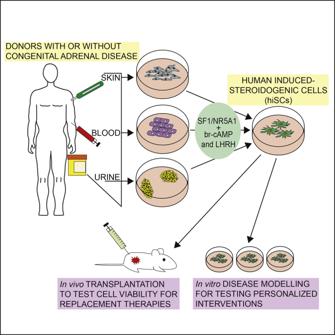
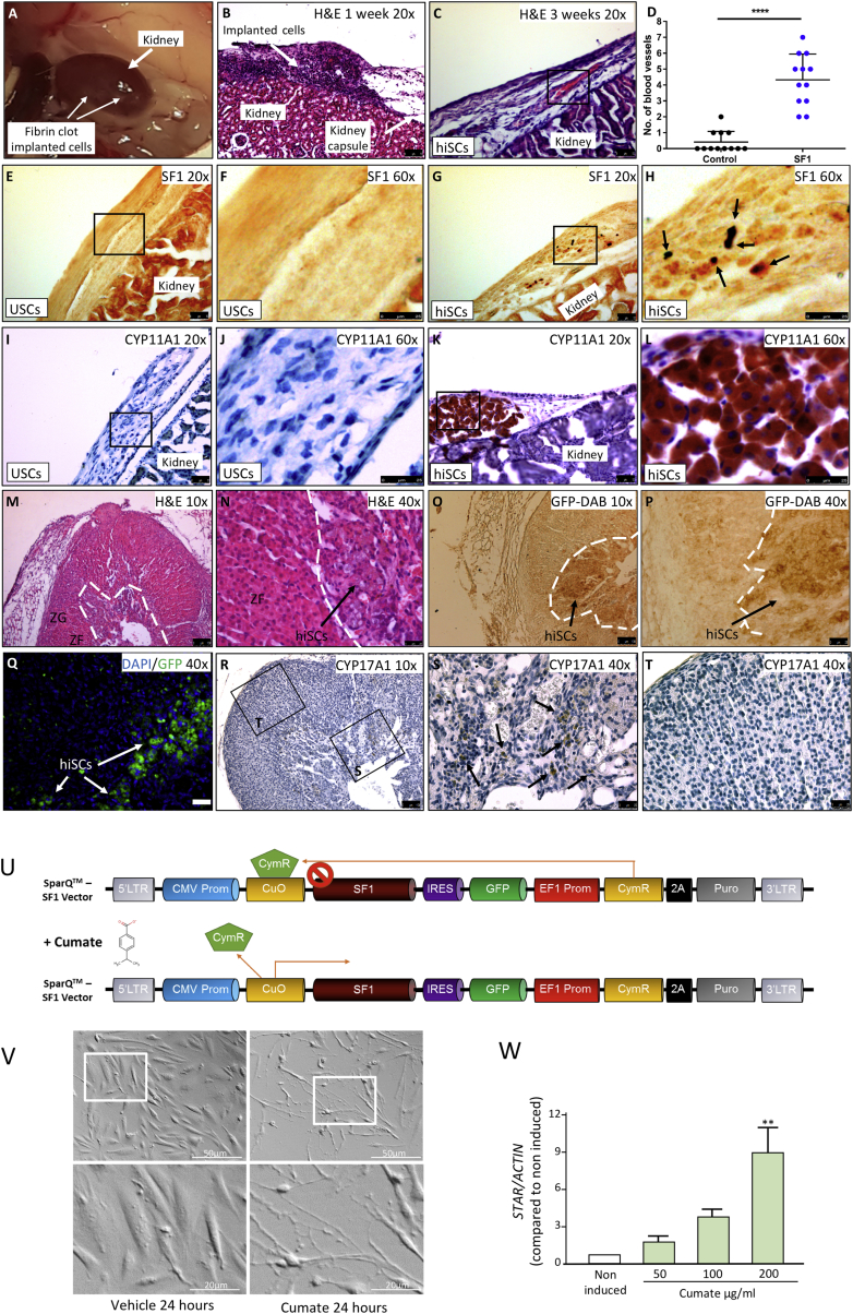
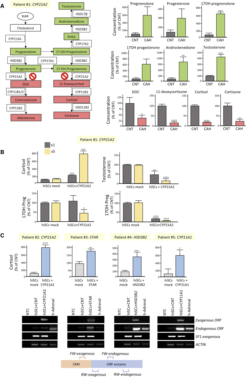
Adrenal insufficiency is managed by hormone replacement therapy, which is far from optimal; the ability to generate functional steroidogenic cells would offer a unique opportunity for a curative approach to restoring the complex feedback regulation of the hypothalamic-pituitary-adrenal axis. Here, we generated human induced steroidogenic cells (hiSCs) from fibroblasts, blood-, and urine-derived cells through forced expression of steroidogenic factor-1 and activation of the PKA and LHRH pathways. hiSCs had ultrastructural features resembling steroid-secreting cells, expressed steroidogenic enzymes, and secreted steroid hormones in response to stimuli. hiSCs were viable when transplanted into the mouse kidney capsule and intra-adrenal. Importantly, the hypocortisolism of hiSCs derived from patients with adrenal insufficiency due to congenital adrenal hyperplasia was rescued by expressing the wild-type version of the defective disease-causing enzymes. Our study provides an effective tool with many potential applications for studying adrenal pathobiology in a personalized manner and opens venues for the development of precision therapies.

Keywords: NR5A1; adrenal cortex; adrenal insufficiency; congenital adrenal hyperplasia; disease modeling; reprogramming; steroidogenic cells; steroidogenic factor 1; transplantation; urine-derived stem cells.


Figures

None
Graphical abstract
Figure 1
Figure 1
Conversion of Human Urine-Derived Stem Cells into Steroidogenic Cells (A) Schematic illustrating our strategy for urine collection, processing, and reprogramming. Urine-derived cells (USCs) were cultured in specific media, and type-II colonies amplified and characterized through flow cytometry. Then they were either banked or expanded for experiments. USCs were infected at passage two with either a lentivirus encoding a transcription factor (TF) within an IRES-GFP vector, a combination of TFs, or mock infected (MOI = 200). Cells were treated with 8-br-cAMP (100 μM) unless stated otherwise and kept in culture for at least eight days before analyses. (B) RT-PCR showing STAR expression on forced expression of each TF. The expression of exogenous SF1, PBX1, WT1, DAX1, and CITED2 was assessed by RT-PCR using primers encompassing the coding- and vector- specific regions. Human adrenal cDNA was used as a positive control for endogenous STAR expression and, along with non-template control (NTC), as a negative control for exogenous TF expression. (C) qRT-PCR analyses of STAR expression on forced expression of SF1 with each TF (upper panel) and of SF1 with or without a combination of TFs (lower panel). (D) Western blot analyses of PCNA and GAPDH expression in hiSCs and mock-reprogrammed USCs from four independent donors eight days after reprogramming (top left panels); cell counting (bottom left panels) and representative images (right panels) of hiSCs obtained from USCs and fibroblasts versus mock-reprogrammed cells. Scale bars, 50 μm. (E) qRT-PCR analyses of STAR expression on forced expression of SF1 with or without the indicated treatments, started the day after infection for seven days. CNT, cells infected with empty control vector. (F) qRT-PCR (upper panel) and RT-PCR (lower panels) analyses of STAR and SF1 expression after reprogramming USCs at different MOI of SF1 or empty control lentiviral vector (CNT). (G) Morphological changes on SF1 overexpression in USCs eight days post-infection. Scale bars, 20 μm. (H) Electron microscopy images of USCs and USCs eight days after reprogramming. Arrows point to mitochondria. Nu, nucleus. Scale bars, 2 μm (left panels) and 1 μm (right panels). Data in (C)–(F) are represented as mean ± SEM, n ≥ 3. See also Figures S1 and S2.
Figure 2
Figure 2
Gene Expression Profile of Reprogrammed USCs (A) Steroidogenic pathway in the adrenal cortex leading to the production of cortisol and aldosterone. (B) RT-PCR expression analyses of STAR, steroidogenic enzymes, SULT2A1, GAPDH, and ACTIN in cells infected with SF1 or control (CNT) lentiviruses after eight days. Human adrenal cDNA was used as a positive control. All cells were treated with 8-br-cAMP. NTC, no template control. (C) Western blot analyses of STAR, SF1, steroidogenic enzymes, SULT2A1, and GAPDH expression in USCs and in USCs infected with SF1 or control lentiviruses (mock) after eight days. Human adrenal lysate was used as a positive control. All cells were treated with 8-br-cAMP. (D) Immunostaining of SF1 (upper panels) and STAR (lower panels) in mock-reprogrammed and reprogrammed USCs. Scale bars, 20 μm; insets, 5 μm. Data in (B) are represented as mean ± SEM, n ≥ 3. See also Figure S3.
Figure 3
Figure 3
Hormone Production in Reprogrammed USCs (A) LC-MS/MS analyses of steroid in the media of reprogrammed versus control USCs after eight days. N.D., non-detected. (B) RT-PCR showing MC2R and MRAP expression in controls and reprogrammed USCs after eight days. (C) Cortisol production in control and hiSCs treated with 1 μM ACTH or 100 μM 8-br-cAMP for eight days. (D) qRT-PCR analyses of STAR, CYP11B1, and CYP11B2 expression in controls and reprogrammed USCs treated with LH and WNT4 for eight days. Cortisol secretion is reported in the right panel. (E) qRT-PCR analyses of STAR expression in USCs treated with LHRH, bombesin, and ACTH. Effect of [D-Trp6]-LHRH and bombesin on cortisol production (left) and in cell viability using CC8 assay. (F) Schematic illustrating the final protocol employed to generate hiSCs. Data in (A) and (C)–(E) are represented as mean ± SEM, n ≥ 3. See also Figure S4.
Figure 4
Figure 4
Transplantation of hiSCs into Mouse Kidney and Adrenal Gland (A) hiSCs implanted directly under the kidney capsule as a fibrin clot. (B–D) H&E analyses of cells directly implanted under the kidney capsule as a fibrin clot. Blood vessels were observed to develop in explants after three weeks when hiSCs were implanted directly under the kidney capsule as a fibrin clot (C, count in D). (E–H) Expression of SF1 in controls (E and F) and hiSCs (G and H) implanted directly under the kidney capsule. (I–L) Expression of CYP11A1 in controls (I and J) and hiSCs (K and L) implanted directly under the kidney capsule. (M and N) H&E analyses of a mouse adrenal gland transplanted for one week with USCs infected with SF1 24 hr earlier at lower (M) and higher (N) magnification. hiSCs can be observed at the cortex/medulla boundary (arrow in N). (G–T) Cells transplanted into the adrenal can be visualized by the expression of GFP with immunohistochemistry (O and P), GFP indirect immunofluorescence (Q), or by their expression of CYP17A1 via immunohistochemistry (R–T). Arrows in (O)–(Q) and (S) point to transplanted cells. CYP17A1 is absent in mouse adrenal cortex (R and T). ZG, zona glomerulosa; ZF, zona fasciculata. Scale bars: 50 μm in (B), (C), (E), (G), (I), and (K); 25 μm in (F), (H), (J), (L), (N), (P), (Q), (S), (T); and 100 μm (M), (O), (R). (U–W) Development of an inducible system aimed at generating unlimited amounts of hiSCs. (U) Schematic of the cumate vector generated to infect USCs. In repressed configuration, CymR repressor strongly binds to the cumate operator site (CuO), downstream of the CMV5 promoter. When cumate is present, CymR is released, which enables transgene expression. (V) USCs were infected with the cumate vector, selected with puromycin, and then treated with cumate or vehicle. Cells underwent similar morphologic changes to those observed in Figure 1G. Scale bars, 50 μm; inset, 20 μm. (W) qRT-PCR analyses of STAR expression after seven days of treatment with increasing concentration of cumate, showing induction dose dependently. Data in (W) are represented as mean ± SEM, n = 3. See also Figure S5.
Figure 5
Figure 5
Characterization of Urine-Derived hiSCs Established from Patients with CAH (A) Comparison of steroidogenic profile of hiSCs established from patient #1 with CYP21A2 mutation (CAH) versus healthy donors (CNT). The diagram on the left shows the steroidogenic pathway with increased metabolites in patient #1 highlighted in green and decreased ones in red. (B) Comparison of cortisol, 17-hydroxyprogesterone, 17-hydroxypregnenolone, and testosterone levels of hiSCs derived from patient #1 with or without restoration of the wild-type form 21-OH. Cells were infected with two increasing amounts of lentiviral particles (×1 and ×5). (C) Comparison of cortisol levels of hiSCs derived from patients (#2–#5) with several forms of CAH with or without restoration of the wild-type form of the corresponding steroidogenic enzymes. RT-PCR analyses using primers encompassing coding- and vector-specific regions confirmed the expression of the exogenous enzymes (lower panels). See also Table 1. Data are represented as mean ± SEM, n ≥ 3.

References

    1. Achermann J.C., Meeks J.J., Jameson J.L. Phenotypic spectrum of mutations in DAX-1 and SF-1. Mol. Cell. Endocrinol. 2001;185:17–25. - PubMed
    1. Andreu Z., Yáñez-Mó M. Tetraspanins in extracellular vesicle formation and function. Front. Immunol. 2014;5:442. - PMC - PubMed
    1. Balyura M., Gelfgat E., Ehrhart-Bornstein M., Ludwig B., Gendler Z., Barkai U., Zimerman B., Rotem A., Block N.L., Schally A.V., Bornstein S.R. Transplantation of bovine adrenocortical cells encapsulated in alginate. Proc. Natl. Acad. Sci. USA. 2015;112:2527–2532. - PMC - PubMed
    1. Bergthorsdottir R., Leonsson-Zachrisson M., Odén A., Johannsson G. Premature mortality in patients with Addison’s disease: a population-based study. J. Clin. Endocrinol. Metab. 2006;91:4849–4853. - PubMed
    1. Bharadwaj S., Liu G., Shi Y., Wu R., Yang B., He T., Fan Y., Lu X., Zhou X., Liu H. Multipotential differentiation of human urine-derived stem cells: potential for therapeutic applications in urology. Stem Cells. 2013;31:1840–1856. - PubMed

Publication types

MeSH terms

Substances