

Age-Associated Decline in Thymic B Cell Expression of Aire and Aire-Dependent Self-Antigens
- PMID: 29386114
- PMCID: PMC5813500
- DOI: 10.1016/j.celrep.2018.01.015
Age-Associated Decline in Thymic B Cell Expression of Aire and Aire-Dependent Self-Antigens
Abstract
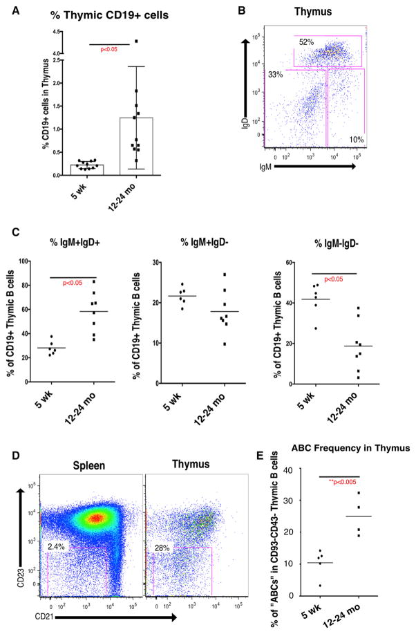
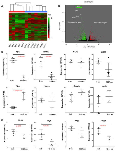
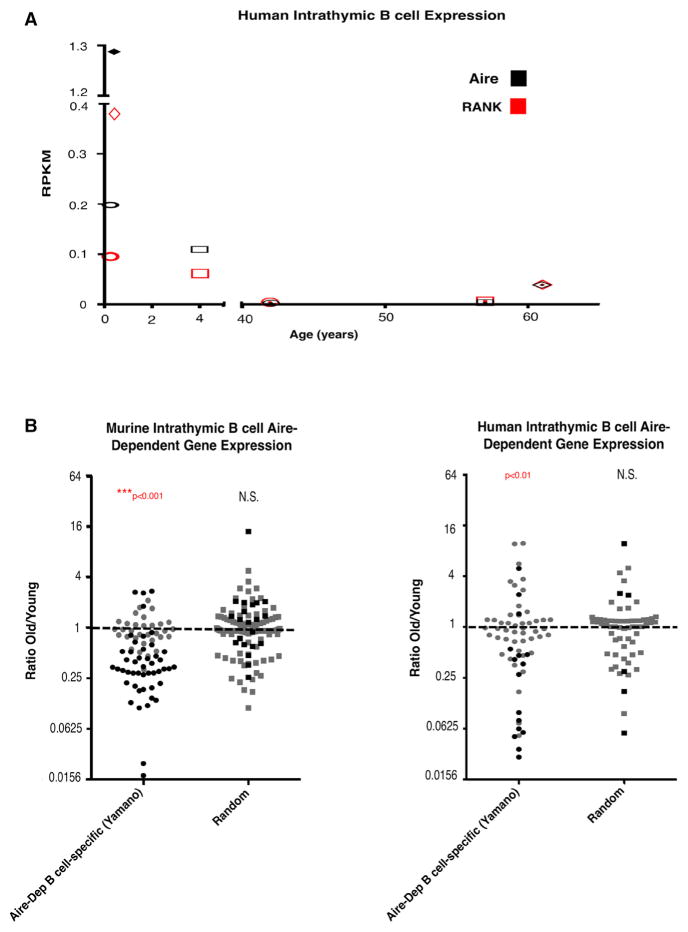
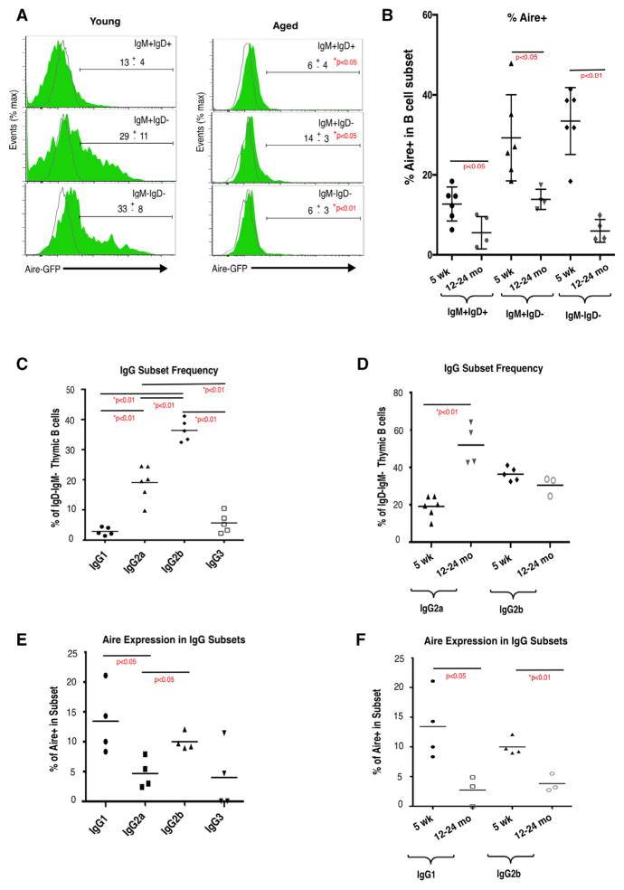
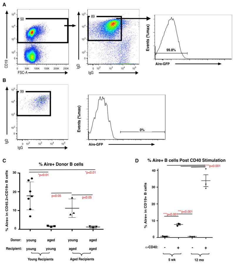
Although autoimmune disorders are a significant source of morbidity and mortality in older individuals, the mechanisms governing age-associated increases in susceptibility remain incompletely understood. Central T cell tolerance is mediated through presentation of self-antigens by cells constituting the thymic microenvironment, including epithelial cells, dendritic cells, and B cells. Medullary thymic epithelial cells (mTECs) and B cells express distinct cohorts of self-antigens, including tissue-restricted self-antigens (TRAs), such that developing T cells are tolerized to antigens from peripheral tissues. We find that expression of the TRA transcriptional regulator Aire, as well as Aire-dependent genes, declines with age in thymic B cells in mice and humans and that cell-intrinsic and cell-extrinsic mechanisms contribute to the diminished capacity of peripheral B cells to express Aire within the thymus. Our findings indicate that aging may diminish the ability of thymic B cells to tolerize T cells, revealing a potential mechanistic link between aging and autoimmunity.
Keywords: Aire; B cell; aging; thymus.
Copyright © 2018 The Author(s). Published by Elsevier Inc. All rights reserved.
Conflict of interest statement
The authors declare no competing interests.
Figures
Similar articles
-
Central T cell tolerance: Identification of tissue-restricted autoantigens in the thymus HLA-DR peptidome.J Autoimmun. 2015 Jun;60:12-9. doi: 10.1016/j.jaut.2015.03.004. Epub 2015 Apr 21. J Autoimmun. 2015. PMID: 25911201
-
Aire mediates thymic expression and tolerance of pancreatic antigens via an unconventional transcriptional mechanism.Eur J Immunol. 2013 Jan;43(1):75-84. doi: 10.1002/eji.201242761. Epub 2012 Dec 6. Eur J Immunol. 2013. PMID: 23041971
-
Aire-dependent thymic expression of desmoglein 3, the autoantigen in pemphigus vulgaris, and its role in T-cell tolerance.J Invest Dermatol. 2011 Feb;131(2):410-7. doi: 10.1038/jid.2010.330. Epub 2010 Nov 4. J Invest Dermatol. 2011. PMID: 21048786
-
Medullary thymic epithelial cells, the indispensable player in central tolerance.Sci China Life Sci. 2013 May;56(5):392-8. doi: 10.1007/s11427-013-4482-4. Epub 2013 May 1. Sci China Life Sci. 2013. PMID: 23633070 Review.
-
Expression of AIRE in thymocytes and peripheral lymphocytes.Autoimmunity. 2008 Mar;41(2):133-9. doi: 10.1080/08916930701773941. Autoimmunity. 2008. PMID: 18324482 Review.
Cited by
-
Early life imprinting of intestinal immune tolerance and tissue homeostasis.Immunol Rev. 2024 May;323(1):303-315. doi: 10.1111/imr.13321. Epub 2024 Mar 19. Immunol Rev. 2024. PMID: 38501766 Free PMC article. Review.
-
The DARC Side of Inflamm-Aging: Duffy Antigen Receptor for Chemokines (DARC/ACKR1) as a Potential Biomarker of Aging, Immunosenescence, and Breast Oncogenesis among High-Risk Subpopulations.Cells. 2022 Nov 29;11(23):3818. doi: 10.3390/cells11233818. Cells. 2022. PMID: 36497078 Free PMC article. Review.
-
Thymic B Cells Promote Germinal Center-Like Structures and the Expansion of Follicular Helper T Cells in Lupus-Prone Mice.Front Immunol. 2020 Apr 28;11:696. doi: 10.3389/fimmu.2020.00696. eCollection 2020. Front Immunol. 2020. PMID: 32411134 Free PMC article.
-
CCR4 and CCR7 differentially regulate thymocyte localization with distinct outcomes for central tolerance.Elife. 2023 Jun 2;12:e80443. doi: 10.7554/eLife.80443. Elife. 2023. PMID: 37266571 Free PMC article.
-
Immunosenescence: a key player in cancer development.J Hematol Oncol. 2020 Nov 10;13(1):151. doi: 10.1186/s13045-020-00986-z. J Hematol Oncol. 2020. PMID: 33168037 Free PMC article. Review.
References
-
- Akashi K, Richie LI, Miyamoto T, Carr WH, Weissman IL. B lymphopoiesis in the thymus. J Immunol. 2000;164:5221–5226. - PubMed
-
- Akiyama T, Shimo Y, Yanai H, Qin J, Ohshima D, Maruyama Y, Asaumi Y, Kitazawa J, Takayanagi H, Penninger JM, et al. The tumor necrosis factor family receptors RANK and CD40 cooperatively establish the thymic medullary microenvironment and self-tolerance. Immunity. 2008;29:423–437. - PubMed
-
- Anderson MS, Venanzi ES, Klein L, Chen Z, Berzins SP, Turley SJ, von Boehmer H, Bronson R, Dierich A, Benoist C, Mathis D. Projection of an immunological self shadow within the thymus by the aire protein. Science. 2002;298:1395–1401. - PubMed
Publication types
MeSH terms
Substances
Grants and funding
LinkOut - more resources
Full Text Sources
Other Literature Sources
Medical
Molecular Biology Databases
