. 2018 Jan 23;22(4):1003-1015.
doi: 10.1016/j.celrep.2017.12.093. Epub 2018 Jan 28.

Epithelial WNT Ligands Are Essential Drivers of Intestinal Stem Cell Activation

Affiliations

Epithelial WNT Ligands Are Essential Drivers of Intestinal Stem Cell Activation

Winnie Y Zou et al. Cell Rep. .

Abstract

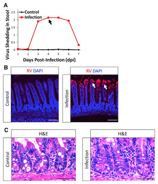
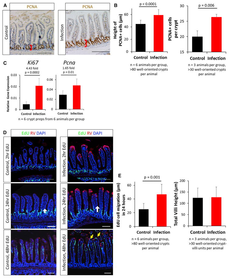
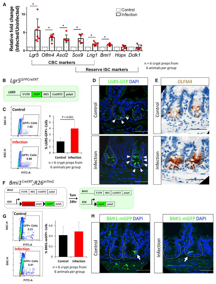
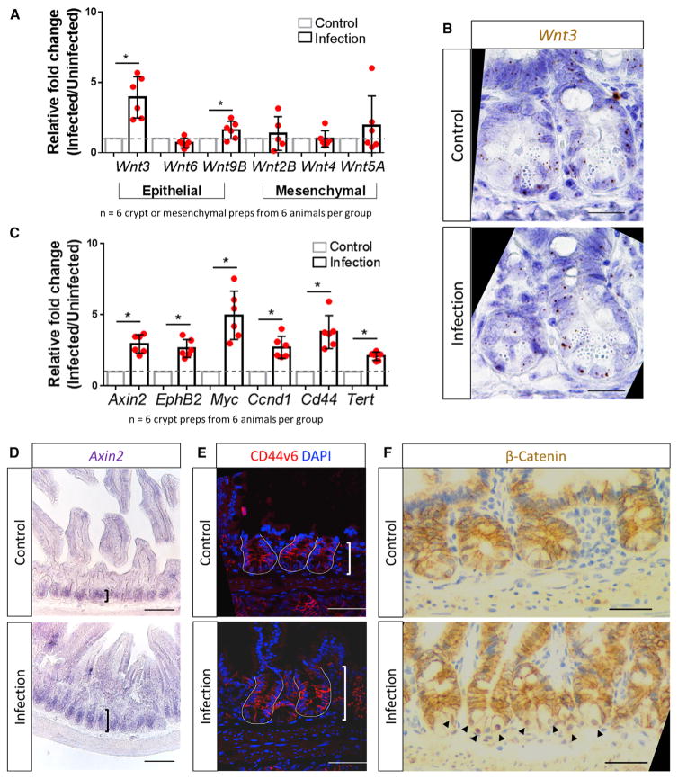
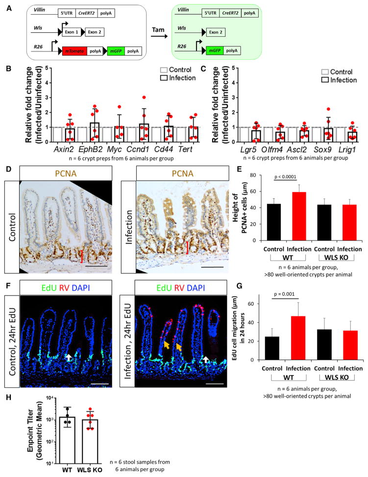
Intestinal stem cells (ISCs) maintain and repair the intestinal epithelium. While regeneration after ISC-targeted damage is increasingly understood, injury-repair mechanisms that direct regeneration following injuries to differentiated cells remain uncharacterized. The enteric pathogen, rotavirus, infects and damages differentiated cells while sparing all ISC populations, thus allowing the unique examination of the response of intact ISC compartments during injury-repair. Upon rotavirus infection in mice, ISC compartments robustly expand and proliferating cells rapidly migrate. Infection results specifically in stimulation of the active crypt-based columnar ISCs, but not alternative reserve ISC populations, as is observed after ISC-targeted damage. Conditional ablation of epithelial WNT secretion diminishes crypt expansion and ISC activation, demonstrating a previously unknown function of epithelial-secreted WNT during injury-repair. These findings indicate a hierarchical preference of crypt-based columnar cells (CBCs) over other potential ISC populations during epithelial restitution and the importance of epithelial-derived signals in regulating ISC behavior.

Keywords: ISC; RV; epithelial WNT; intestinal stem cell; regeneration; rotavirus.


Conflict of interest statement

DECLARATION OF INTERESTS

The authors declare no competing interests.

Figures

Figure 1
Figure 1. RV Infection Is Limited to the Tip of the Villi, Preserving an Intact Crypt Compartment
(A) Representative stool ELISA monitoring RV infections. Points represent means of respective groups (n = 2). Peak viral shedding was observed at 4 days post-infection (dpi; black arrow). (B) Representative confocal images of control- and RV-infected mouse epithelium. RV-infected differentiated cells were detected using a laboratory-generated, polyclonal anti-RV antibody. Infected villi are noted by white arrows. (C) Representative H&E images showing intact crypts in both control- and RV-infected mouse epithelium. See also Figures S1 and S2.
Figure 2
Figure 2. RV Infection Led to Increased Epithelial Proliferation and Increased Migration
(A) Representative immunohistochemistry images of PCNA+ proliferating compartments. Red brackets represent the height of the PCNA+ cells measured in (B). (B) Quantification of height of PCNA+ cells (left) and PCNA+ cells per crypt (right). (C) qRT-PCR results showing upregulation of the proliferative markers Ki67 and Pcna in isolated epithelial crypts. (D) Representative confocal images of cell migration in control- and RV-infected animals with 2-, 24-, and 48-hr EdU labeling. EdU+ cells were restricted to the crypts after 2-hr EdU labeling. The length of 24-hr EdU migration was measured from the crypt-villi junction to the farthest EdU-labeled cells (white arrows). After 48 hr of labeling, EdU+ cells were observed near RV-infected cells (yellow arrows). (E) Quantification of cell migration 24 hr after EdU labeling (left) and total villi height (right). EdU cell migration distance is an absolute measure of distance from the crypt-villus junction (just outside of the transient amplifying [TA] zone) to the cell that had migrated the furthest. Bars represent means ± SD. All statistical analyses were performed using Student’s t test. Scale bars, 100 μm. See also Figures S3–S6.
Figure 3
Figure 3. CBCs Are Induced following RV Infection
(A) qRT-PCR results showed upregulation of all CBC markers and some reserve ISC markers. (B) Schematic of Lgr5GFPCreERT mice. Cells expressing LGR5 are green. (C) Representative flow cytometry analysis on control- and RV-infected Lgr5GFPCreERTmice. Quantification of GFP+ cells using flow cytometry analysis on crypt-enriched epithelial preparations. (D) Representative confocal images of Lgr5GFPCreERT mice following control- and RV-infection. White arrowheads denote GFP+ cells. (E) Representative light microscopy images of OLFM4 immunohistochemistry staining in control- and RV-infected animals. (F) Schematic of Bmi1CreERT;R26mTmG mice. (G) Representative flow cytometry analysis on control- and RV-infected Bmi1CreERT;R26mTmG mice. Quantification of BMI1-mGFP+ cells using flow cytometry analysis on crypt-enriched epithelial preparations. (H) Representative confocal images of BMI1-mGFP+ mice following control- and RV-infection. White arrows denote BMI1-mGFP+ cells. Scale bars, 50 μm.
Figure 4
Figure 4. WNT Signaling Pathway Is Stimulated in Crypts following RV Infection
(A) qRT-PCR results showing upregulation of the epithelial-expressed Wnt3 and Wnt9B in isolated epithelial crypt preparations. Wnt2B, Wnt4, and Wnt5A remained stable in mesenchymal preparations. (B) RNAScope analysis showing Wnt3 expression in Paneth cells and other crypt cell types in both control and RV infection. (C) qRT-PCR results showing upregulation of well-established WNT signaling pathway target genes in isolated epithelial crypt preparations after RV infection. (D) Representative RNA in situ hybridization images showed expansion of Axin2 expression in RV infected animals. (E) Representative immunofluorescence images showing that the WNT-target gene CD44v6 is expressed on more cells in the RV-infected animal. (F) Representative immunohistochemistry staining of β-catenin, the intracellular transducer of the WNT signaling pathway. RV infection induces cytoplasm to nuclear translocation of β-catenin in the crypts (black arrowheads). Scale bars represent 50 μm in (B), (E), and (F) and 100 μm in (D). See also Figure S6.
Figure 5
Figure 5. Epithelial-Secreted WNT Ligands Are Essential for RV-Induced Stem Cell Regeneration
(A) Schematic of VillinCreERT;WLSf/f;R26mTmG (WLS KO) mouse. Injections of tamoxifen allows for the conditional knockout of the Wntless gene in villin-expressing cells, impairing WNT secretion specifically in the epithelium. (B) qRT-PCR results showed expression of WNT pathway target genes remain stable in isolated epithelial crypts following RV infection in WLS KO mice. (C) qRT-PCR results showed expression of putative CBC markers remain stable in isolated epithelial crypts following RV infection in WLS KO mice. (D) Representative immunohistochemistry staining of PCNA in control- and RV-infected WLS KO animals. (E) Quantification of height of PCNA+ cell measurement in WT and WLS KO mice. Bars represent means ± SD. (F) Representative image of EdU-labeled cell migration in control- and RV-infected animals in WLS KO mice. There was no induction of EdU+ cell migration (white arrows). RV-infected cells were no longer restricted to the tip of the villi (orange arrows). (G) Quantification of cell migration with 24 hr of EdU labeling in WT and WLS KO mice (white arrows in E). Bars represent means ± SD. (H) Endpoint titer from stool ELISA of WT and WLS KO animals. Bars represent geometric mean ± 95% CI. Scale bars, 100 μm. See also Figure S7.

References

    1. Aoki R, Shoshkes-Carmel M, Gao N, Shin S, May CL, Golson ML, Zahm AM, Ray M, Wiser CL, Wright CV, Kaestner KH. Foxl1-expressing mesenchymal cells constitute the intestinal stem cell niche. Cell Mol Gastroenterol Hepatol. 2016;2:175–188. - PMC - PubMed
    1. Arnold MM, Sen A, Greenberg HB, Patton JT. The battle between rotavirus and its host for control of the interferon signaling pathway. PLoS Pathog. 2013;9:e1003064. - PMC - PubMed
    1. Bänziger C, Soldini D, Schütt C, Zipperlen P, Hausmann G, Basler K. Wntless, a conserved membrane protein dedicated to the secretion of Wnt proteins from signaling cells. Cell. 2006;125:509–522. - PubMed
    1. Barker N, van Es JH, Kuipers J, Kujala P, van den Born M, Cozijnsen M, Haegebarth A, Korving J, Begthel H, Peters PJ, Clevers H. Identification of stem cells in small intestine and colon by marker gene Lgr5. Nature. 2007;449:1003–1007. - PubMed
    1. Beau I, Berger A, Servin AL. Rotavirus impairs the biosynthesis of brush-border-associated dipeptidyl peptidase IV in human enterocyte-like Caco-2/TC7 cells. Cell Microbiol. 2007;9:779–789. - PubMed

Publication types