. 2018 Mar 16;122(6):836-845.
doi: 10.1161/CIRCRESAHA.118.312660. Epub 2018 Jan 31.

Enhanced Redox State and Efficiency of Glucose Oxidation With miR Based Suppression of Maladaptive NADPH-Dependent Malic Enzyme 1 Expression in Hypertrophied Hearts

Affiliations

Enhanced Redox State and Efficiency of Glucose Oxidation With miR Based Suppression of Maladaptive NADPH-Dependent Malic Enzyme 1 Expression in Hypertrophied Hearts

Ryan Lahey et al. Circ Res. .

Abstract

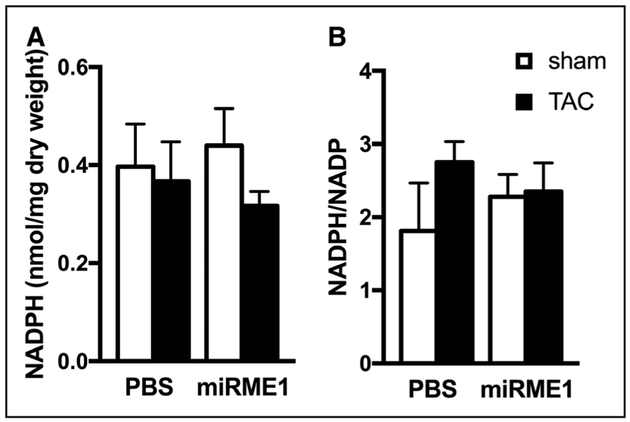
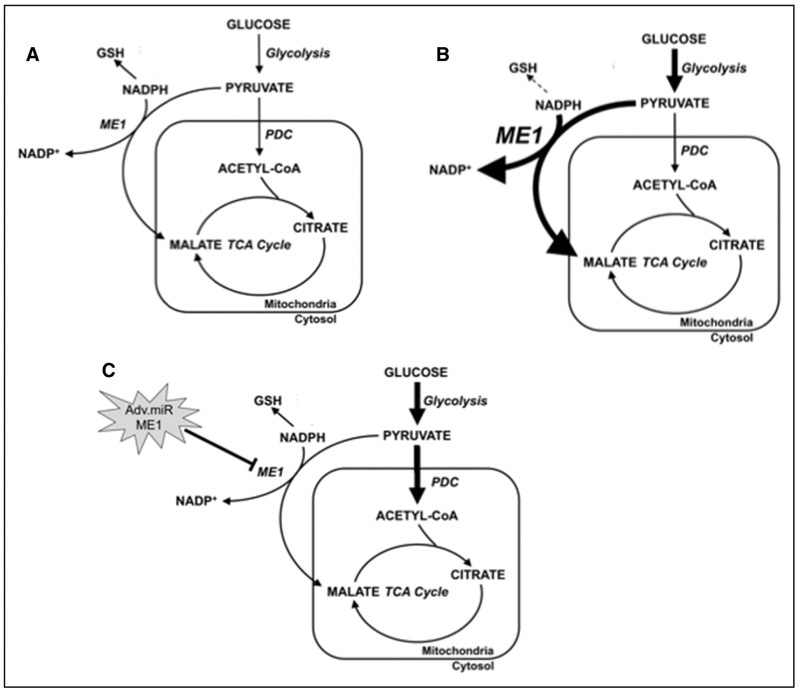
Rationale: Metabolic remodeling in hypertrophic hearts includes inefficient glucose oxidation via increased anaplerosis fueled by pyruvate carboxylation. Pyruvate carboxylation to malate through elevated ME1 (malic enzyme 1) consumes NADPH necessary for reduction of glutathione and maintenance of intracellular redox state.

Objective: To elucidate upregulated ME1 as a potential maladaptive mechanism for inefficient glucose oxidation and compromised redox state in hypertrophied hearts.

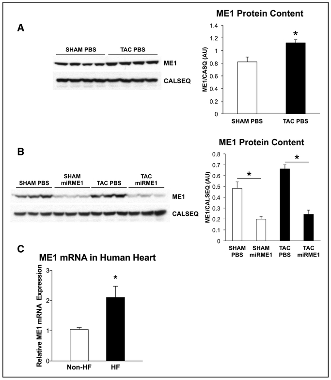
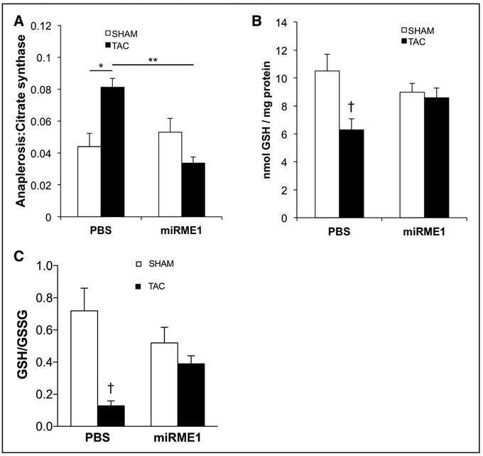
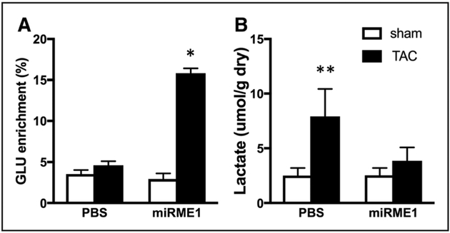
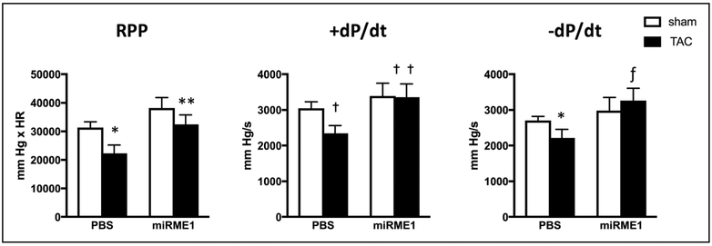
Methods and results: ME1 expression was selectively inhibited, in vivo, via non-native miR-ME1 (miRNA specific to ME1) in pressure-overloaded rat hearts. Rats subjected to transverse aortic constriction (TAC) or Sham surgery received either miR-ME1 or PBS. Effects of ME1 suppression on anaplerosis and reduced glutathione (GSH) content were studied in isolated hearts supplied 13C-enriched substrate: palmitate, glucose, and lactate. Human myocardium collected from failing and nonfailing hearts during surgery enabled RT-qPCR confirmation of elevated ME1 gene expression in clinical heart failure versus nonfailing human hearts (P<0.04). TAC induced elevated ME1 content, but ME1 was lowered in hearts infused with miR-ME1 versus PBS. Although Sham miR-ME1 hearts showed no further reduction of inherently low anaplerosis in normal heart, miR-ME1 reduced anaplerosis in TAC to baseline: TAC miR-ME1=0.034±0.004; TAC PBS=0.081±0.005 (P<0.01). Countering elevated anaplerosis in TAC shifted pyruvate toward oxidation in the tricarboxylic acid cycle. Importantly, via the link to NADPH consumption by pyruvate carboxylation, ME1 suppression in TAC restored GSH content, reduced lactate production, and ultimately improved contractility.

Conclusions: A maladaptive increase in anaplerosis via ME1 in TAC is associated with reduced GSH content. Suppressing increased ME1 expression in hypertrophied rat hearts, which is also elevated in failing human hearts, reduced pyruvate carboxylation thereby normalizing anaplerosis, restoring GSH content, and reducing lactate accumulation. Reducing ME1 induced favorable metabolic shifts for carbohydrate oxidation, improving intracellular redox state and enhanced cardiac performance in pathological hypertrophy.

Keywords: glucose; heart failure; hypertrophy; metabolism; microRNAs.


Figures

Figure 1.
Figure 1.. Protein levels of ME1 (malic enzyme 1) in pressure-overloaded rat hearts and ME1 message levels in failing human hearts.
A, Protein expression of ME1 was increased in transverse aortic constriction (TAC) PBS compared with sham-operated (SHAM) PBS (n=4 for both groups). B, Delivery of adenovirus containing miR-ME1 (miRNA specific to ME1) vector reduced protein expression of ME1 in both groups (n=3 for each group). White bar, PBS injected; black bar, miR-ME1 injected. *P<0.01. Graphs represent the quantitative analysis of each corresponding blot using NIH ImageJ software. C, mRNA levels of the gene encoding ME1 were elevated in failing human hearts (HF) versus that of nonfailing human hearts (non-HF). *P<0.02 with Students t test and P<0.04 with Welch t test. CALSEQ indicates calsequestrin
Figure 2.
Figure 2.. Anaplerosis and reduced glutathione content (GSH) in transverse aortic constriction (TAC) hearts.
A, Anaplerosis, expressed as a ratio to citrate synthase activity. In the PBS group, anaplerotic flux was increased nearly 100% with TAC. Knockdown of ME1 expression significantly reduced anaplerosis in TAC to normal levels. *P<0.01;**P<0.001 by ANOVA and Tukey–Kramer test. B, Compared with sham operated (SHAM), TAC hearts demonstrated significantly lower GSH content, as expected. Suppression of ME1 (malic enzyme 1) expression (miRME1 [miRNA specific to ME1]) restored GSH content in TAC to levels comparable to SHAM. C, GSH/GSSG ratios were also improved in the miR-ME1 TAC group in comparison to reduced GSH/GSSG in the Sham-infused TAC hearts. White bar, SHAM; black bar, TAC. †P<0.05 vs SHAM PBS by ANOVA and Tukey–Kramer test.
Figure 3.
Figure 3.. NADPH (A) and NADPH/NADP (B) ratio were not significantly affected in response to ME1 (malic enzyme 1) suppression.
Consumption of NADPH to maintain reduced glutathione (GSH) levels in transverse aortic constriction (TAC) hearts with ME1 suppression is reflected in lack of differences in the values among experimental groups. White bars, Sham operated (SHAM); Black bars, TAC. miR-ME1 indicates miRNA specific to ME1.
Figure 4.
Figure 4.. Increased carbohydrate oxidation in hearts with miR ME1 (miRNA specific to malic enzyme 1) suppression and attenuated lactate accumulation after transverse aortic constriction (TAC).
A, Glutamate (GLU) 13C enrichment from 13C glucose and 13C lactate reflecting oxidation of pyruvate from glycolysis and lactate. Note increased isotopic enrichment of GLU in TAC hearts receiving miR-ME1 to suppress pyruvate carboxylation and enhance oxidation via pyruvate decarboxylase. B, Tissue lactate levels. ME1 suppression prevented the accumulation of lactate in TAC hearts. White bars, Sham operated (SHAM); Black bars, TAC. *P<0.05 vs all other experimental groups; **P<0.05 vs sham hearts by ANOVA and Tukey–Kramer test.
Figure 5.
Figure 5.. Contractile response to ME1 (malic enzyme 1) suppression in Sham operated (Sham) and transverse aortic constriction (TAC) hearts.
Direct comparison between mean values reveals improved dP/dt in TAC hearts receiving miR-ME1 (miRNA specific to ME1) vs TAC. White bars, SHAM; Black bars, TAC. *P<0.05, Sham ME1 vs TAC PBS by ANOVA and Tukey–Kramer test; **P<0.05, TAC PBS vs TAC miR-ME1 by Student t test; †P<0.05, Sham PBS vs TAC PBS by ANOVA and Tukey–Kramer test; ††P=0.05, TAC PBS vs TAC miRME1 by Student t test; ƒP<0.05 TAC PBS vs TAC miR-ME1 by Student t test. RPP indicates rate pressure product.
Figure 6.
Figure 6.. Summary scheme depicting the effects of ME1 (malic enzyme 1) knockdown on glucose and pyruvate metabolism in hypertrophied hearts.
Metabolic fate of glucose and glycolytic end products in a (A) normal heart; (B) pathological heart with increased carbon flux through glycolysis and ME1-mediated entry of carbon into transverse aortic constriction (TCA) cycle via anaplerosis; (C) failing heart treated with virus that suppresses ME1 expression. Increased carbon flux into TCA cycle via pyruvate dehydrogenase complex (PDC) and restoring NADPH-mediated glutathione reduction. Increased metabolic activity is represented by thick arrows. Increased ME1 expression is represented by enlarged text. Decreased reduced glutathione (GSH) formation is represented by dashed line. Adv.miR-ME1 indicates adenovirus containing miR-ME1.

References

    1. Carley AN, Taegtmeyer H, Lewandowski ED. Matrix revisited: mechanisms linking energy substrate metabolism to the function of the heart. Circ Res. 2014;114:717–729. - PMC - PubMed
    1. Doenst T, Nguyen TD, Abel ED. Cardiac metabolism in heart failure: implications beyond ATP production. Circ Res. 2013;113:709–724. - PMC - PubMed
    1. Kolwicz SC Jr, Purohit S, Tian R. Cardiac metabolism and its interactions with contraction, growth, and survival of cardiomyocytes. Circ Res. 2013;113:603–616. doi: 10.1161/CIRCRESAHA.113.302095. - DOI - PMC - PubMed
    1. Ingwall JS, Weiss RG. Is the failing heart energy starved? On using chemical energy to support cardiac function. Circ Res. 2004;95:135–145. doi: 10.1161/01.RES.0000137170.41939.d9. - DOI - PubMed
    1. Lydell CP, Chan A, Wambolt RB, Sambandam N, Parsons H, Bondy GP, Rodrigues B, Popov KM, Harris RA, Brownsey RW, Allard MF. Pyruvate dehydrogenase and the regulation of glucose oxidation in hypertrophied rat hearts. Cardiovasc Res. 2002;53:841–851. - PMC - PubMed

Publication types