. 2018 Feb 13;2(3):210-223.
doi: 10.1182/bloodadvances.2017011254.

Improving T-cell expansion and function for adoptive T-cell therapy using ex vivo treatment with PI3Kδ inhibitors and VIP antagonists

Affiliations

Improving T-cell expansion and function for adoptive T-cell therapy using ex vivo treatment with PI3Kδ inhibitors and VIP antagonists

Christopher T Petersen et al. Blood Adv. .

Abstract

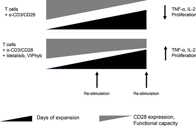
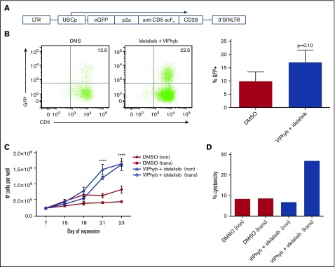
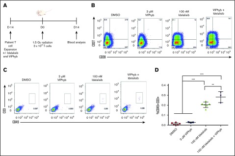
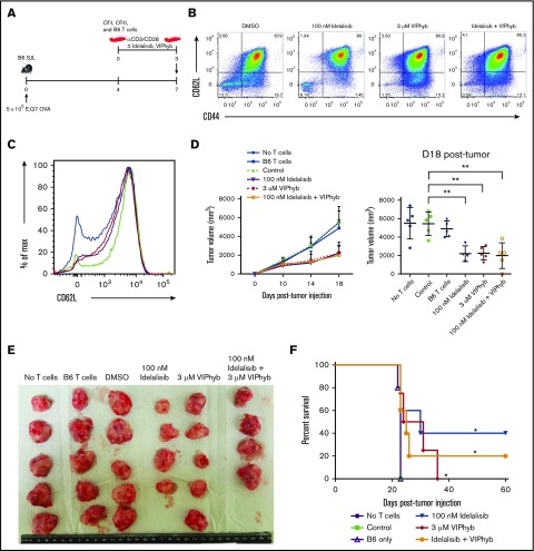
Adoptive therapy with ex vivo-expanded genetically modified antigen-specific T cells can induce remissions in patients with relapsed/refractory cancer. The clinical success of this therapy depends upon efficient transduction and expansion of T cells ex vivo and their homing, persistence and cytotoxicity following reinfusion. Lower rates of ex vivo expansion and clinical response using anti-CD19 chimeric antigen receptor (CAR) T cells have been seen in heavily pretreated lymphoma patients compared with B-cell acute lymphoblastic leukemia patients and motivate the development of novel strategies to enhance ex vivo T cell expansion and their persistence in vivo. We demonstrate that inhibition of phosphatidylinositol 3-kinase δ (PI3Kδ) and antagonism of vasoactive intestinal peptide (VIP) signaling partially inhibits the terminal differentiation of T cells during anti-CD3/CD28 bead-mediated expansion (mean, 54.4% CD27+CD28+ T cells vs 27.4% in control cultures; P < .05). This strategy results in a mean of 83.7% more T cells cultured from lymphoma patients in the presence of PI3Kδ and VIP antagonists, increased survival of human T cells from a lymphoma patient in a murine xenograft model, enhanced cytotoxic activity of antigen-specific human CAR T cells and murine T cells against lymphoma, and increased transduction and expansion of anti-CD5 human CAR T cells. PI3Kδ and VIP antagonist-expanded T cells from lymphoma patients show reduced terminal differentiation, enhanced polyfunctional cytokine expression, and preservation of costimulatory molecule expression. Taken together, synergistic blockade of these pathways is an attractive strategy to enhance the expansion and functional capacity of ex vivo-expanded cancer-specific T cells.


Conflict of interest statement

Conflict-of-interest disclosure: The authors declare no competing financial interests.

Figures

None
Graphical abstract
Figure 1.
Figure 1.
T cells from DLBCL patients who have received multiple rounds of chemotherapy show loss of CD27 and CD28 expression. Peripheral blood samples from healthy controls, untreated DLBCL patients, and heavily-treated DLBCL patients were examined for the expression of CD27 and CD28 by flow cytometry. (A) Representative flow plots of CD27 and CD28 expression gated on CD3+ cells. (B) Quantification of CD27CD28 cells from healthy controls (n = 6), untreated DLBCL patients (n = 5), and treated DLBCL patients (n = 12). Horizontal bar shows mean values; vertical “whiskers” show 95% confidence interval. (C) Linear regression showing the correlation between number of total chemotherapy cycles and frequency of CD27CD28 cells in DLBCL patient samples. Colored symbols and connecting lines indicate paired blood samples taken from DLBCL prior to and following cytotoxic chemotherapy.
Figure 2.
Figure 2.
Depletion of CD27CD28cells improves the expansion of T cells from heavily pretreated DLBCL patients. PBMCs from pretreated DLBCL patients were rested overnight followed by FACS sorting to separate CD27CD28 cells from the remaining populations. Cells were then stimulated with anti-CD3/CD28 beads with IL-2 for 14 days. (A) Comparison of normal T-cell expansion from healthy donors, untreated DLBCL patients, and treated DLBCL patients. (B) Flow plots showing the sorting strategy and postsort purity. (C) T-cell viability at the end of the expansion period as assessed by Sytox blue staining. (D) Quantitation of cell viabilities for the indicated populations on day 14 of expansion (n = 4 DLBCL patient donors). (E) Cell counts of cultures consisting of the indicated T-cell populations from 4 patient sorts. **P < .01, ****P < .001.
Figure 3.
Figure 3.
Pharmacological blockade of PI3K δ and VIP signaling results in increased cell yield, preservation of CD27+CD28+cells and enhanced IL-2 production during ex vivo T-cell expansion. T cells from healthy donors and DLBCL patients were expanded in vitro in the presence or absence of idelalisib and/or VIPhyb for 14 days. Cells were counted on days 7 and 14 of expansion and phenotyped by flow cytometry for surface markers or cytokine production on day 14. (A) Expansion profiles of healthy controls under the indicated culture conditions. (B) Waterfall plot showing the expansion of DLBCL patient samples relative to DMSO controls. The numbers below the bars indicate specific patient samples. (C) Representative flow plots showing the expression of CD27 and CD28 on the total T-cell population. (D) Quantification of double-positive and double-negative T cells from 10 lymphoma patients. (E) Representative flow plots of IL-2 and IFN-γ production by human CD4 and CD8 T cells after 14 days of expansion under the indicated culture conditions. (F) Quantitation of total frequencies of cells producing the indicated cytokines as well as single, double, and triple cytokine-producing T cells (n = 6 healthy donors). *P < .05, **P < .01, ***P < .001. TNF, tumor necrosis factor.
Figure 4.
Figure 4.
Idelalisib and VIPhyb enhance the expansion and functionality of CAR T cells in vitro. CD5 CAR T cells were produced from healthy donors (n = 3) in the presence or absence of idelalisib and VIPhyb and evaluated for growth, transduction efficiency, and in vitro cytotoxicity. (A) CD5 CAR vector design. (B) Transduction efficiency as assessed by GFP expression. (C) Expansion profiles of CD5 CAR T cells under the indicated culture conditions. (D) In vitro cytotoxicity of CD5 CAR T cells generated from healthy controls and expanded under the indicated conditions. eGFP, enhanced green protein; LTR, long terminal repeat; SIN, self-inactivating.
Figure 5.
Figure 5.
Expansion of DLBCL patient T cells in the presence of idelalisib and VIPhyb significantly enhances their in vivo persistence following adoptive transfer to NSG mice. T cells from a heavily treated DLBCL patient were expanded in the presence or absence of idelalisib and/or VIPhyb for 14 days followed by transfer to irradiated NSG mice. Blood was analyzed 14 days posttransfer for the presence of human CD45+CD3+ cells. (A) Experimental outline. (B) Flow plots showing the phenotype of T cells on day 14 of expansion just prior to adoptive transfer. (C) Representative flow plots from the blood of NSG mice showing the frequency of human T cells. (D) Quantification of T-cell frequencies in NSG mice 14 days after adoptive transfer from 1 of 3 independent experiments. **P < .01; ***P < .001.
Figure 6.
Figure 6.
Expansion of T cells in the presence of idelalisib and/or VIPhyb significantly enhances their anti-tumor activity in a murine lymphoma model. OT-I, OT-II, and B6 T cells were expanded for 3 days using anti-CD3/CD28 beads in the presence or absence of idelalisib and/or VIPhyb. Cells were then mixed (2 OT-I:1 OT-II:2 B6 ratio) and injected into B6 SJL mice bearing subcutaneous E.G7 OVA tumors. Growth of tumors was monitored by caliper measurement with volume calculated as (length × width2)/2. (A) Experimental outline. (B) Flow plots showing the phenotype of T cells on day 3 of expansion just prior to injection into tumor-bearing mice. (C) Histogram showing the expression of CD62L on T cells on day 3 of expansion. Blue, DMSO; green, 100 nM idelalisib; purple, 3 μM VIPhyb; red, Idelalisib + VIPhyb. (D) Tumor growth curve and quantification of tumor volume on the final day of allowable growth. (E) Images of tumors removed from sacrificed mice on day 18 of growth. Note that the tumor from 1 mouse from the idelalisib + VIPhyb group regressed and was undetectable. (F) Survival curve of tumor-bearing mice receiving T cells expanded under the indicated conditions. **P < .01; n = 4 or 5 mice per group from 2 independent experiments.

References

    1. Brentjens RJ, Davila ML, Riviere I, et al. . CD19-targeted T cells rapidly induce molecular remissions in adults with chemotherapy-refractory acute lymphoblastic leukemia. Sci Transl Med. 2013;5(177):177ra38. - PMC - PubMed
    1. Martelli M, Ferreri AJ, Agostinelli C, Di Rocco A, Pfreundschuh M, Pileri SA. Diffuse large B-cell lymphoma. Crit Rev Oncol Hematol. 2013;87(2):146-171. - PubMed
    1. Locke FL, Neelapu SS, Bartlett NL, et al. . Phase 1 Results of ZUMA-1: A Multicenter Study of KTE-C19 Anti-CD19 CAR T Cell Therapy in Refractory Aggressive Lymphoma. Mol Ther. 2017;25(1):285-295. - PMC - PubMed
    1. Mackall CL. T-cell immunodeficiency following cytotoxic antineoplastic therapy: a review. Stem Cells. 2000;18(1):10-18. - PubMed
    1. Klebanoff CA, Scott CD, Leonardi AJ, et al. . Memory T cell-driven differentiation of naive cells impairs adoptive immunotherapy. J Clin Invest. 2016;126(1):318-334. - PMC - PubMed

Publication types

MeSH terms