. 2018 Jun 5;137(23):2478-2493.
doi: 10.1161/CIRCULATIONAHA.117.033144. Epub 2018 Jan 31.

CD301b/MGL2+ Mononuclear Phagocytes Orchestrate Autoimmune Cardiac Valve Inflammation and Fibrosis

Affiliations

CD301b/MGL2+ Mononuclear Phagocytes Orchestrate Autoimmune Cardiac Valve Inflammation and Fibrosis

Lee A Meier et al. Circulation. .

Abstract

Background: Valvular heart disease is common and affects the mitral valve (MV) most frequently. Despite the prevalence of MV disease (MVD), the cellular and molecular pathways that initiate and perpetuate it are not well understood.

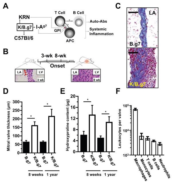
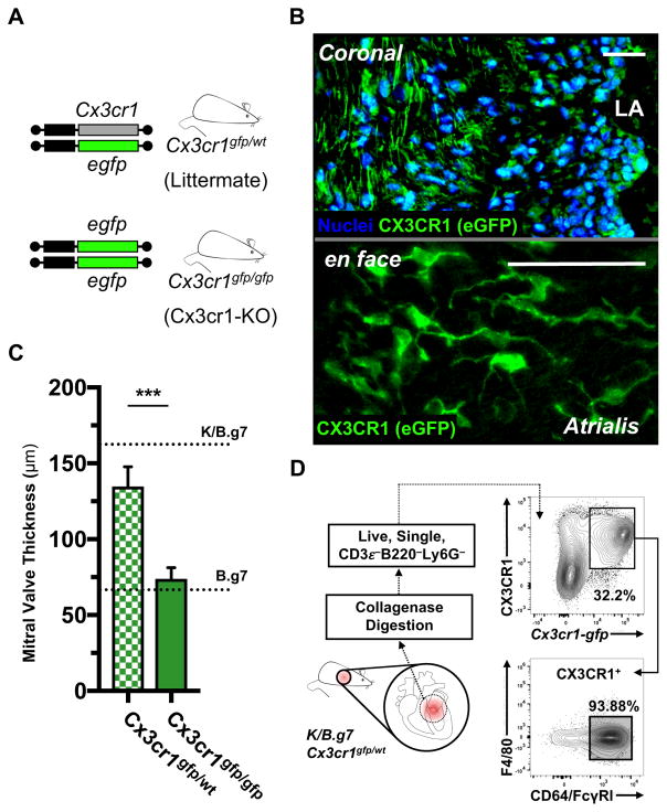
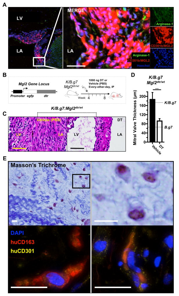
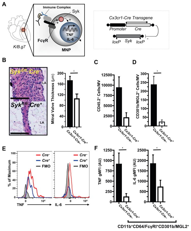
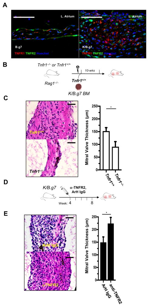
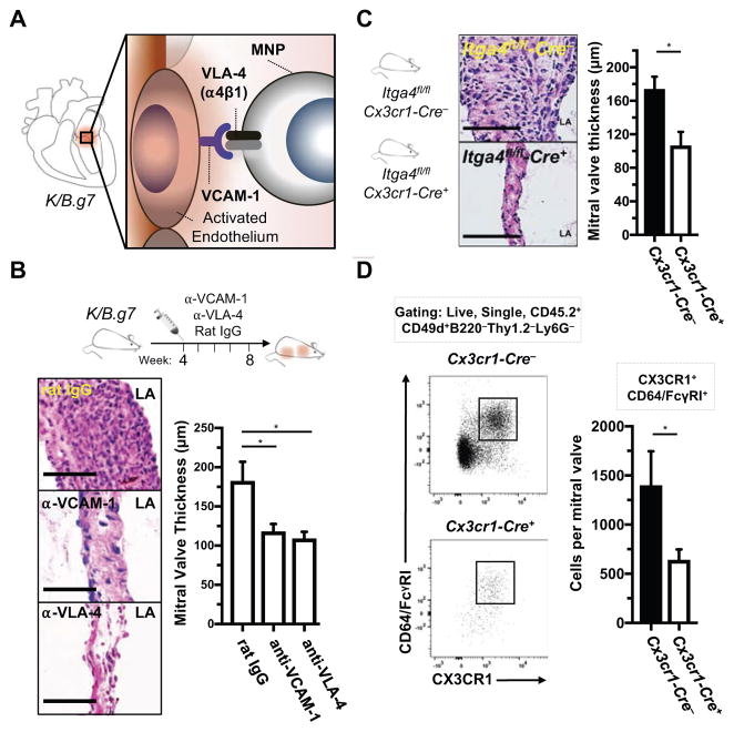
Methods: K/B.g7 T-cell receptor transgenic mice spontaneously develop systemic autoantibody-associated autoimmunity, leading to fully penetrant fibroinflammatory MVD and arthritis. We used multiparameter flow cytometry, intracellular cytokine staining, and immunofluorescent staining to characterize the cells in inflamed K/B.g7 MVs. We used genetic approaches to study the contribution of mononuclear phagocytes (MNPs) to MVD in this model. Specifically, we generated K/B.g7 mice in which either CX3CR1 or CD301b/macrophage galactose N-acetylgalactosamine-specific lectin 2 (MGL2)-expressing MNPs were ablated. Using K/B.g7 mice expressing Cx3Cr1-Cre, we conditionally deleted critical inflammatory molecules from MNPs, including the Fc-receptor signal-transducing tyrosine kinase Syk and the cell adhesion molecule very late antigen-4. We performed complementary studies using monoclonal antibodies to block key inflammatory molecules. We generated bone marrow chimeric mice to define the origin of the inflammatory cells present in the MV and to determine which valve cells respond to the proinflammatory cytokine tumor necrosis factor (TNF). Finally, we examined specimens from patients with rheumatic heart disease to correlate our findings to human pathology.

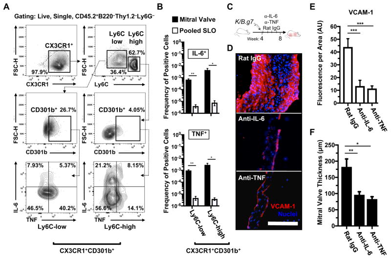
Results: MNPs comprised the vast majority of MV-infiltrating cells; these MNPs expressed CX3CR1 and CD301b/MGL2. Analogous cells were present in human rheumatic heart disease valves. K/B.g7 mice lacking CX3CR1 or in which CD301b/MGL2-expressing MNPs were ablated were protected from MVD. The valve-infiltrating CD301b/MGL2+ MNPs expressed tissue-reparative molecules including arginase-1 and resistin-like molecule α. These MNPs also expressed the proinflammatory cytokines TNF and interleukin-6, and antibody blockade of these cytokines prevented MVD. Deleting Syk from CX3CR1-expressing MNPs reduced their TNF and interleukin-6 production and also prevented MVD. TNF acted through TNF receptor-1 expressed on valve-resident cells to increase the expression of vascular cell adhesion molecule-1. Conditionally deleting the vascular cell adhesion molecule-1 ligand very late antigen-4 from CX3CR1-expressing MNPs prevented MVD.

Conclusions: CD301b/MGL2+ MNPs are key drivers of autoimmune MVD in K/B.g7 mice and are also present in human rheumatic heart disease. We define key inflammatory molecules that drive MVD in this model, including Syk, TNF, interleukin-6, very late antigen-4, and vascular cell adhesion molecule-1.

Keywords: autoimmune diseases; fibrosis; heart valves; inflammation; macrophages; rheumatic heart disease.


Figures

Figure 1
Figure 1. Fully-penetrant, fibro-inflammatory cardiac valve pathology in K/B.g7 mice
A, K/B.g7 mice develop systemic inflammation and auto-antibody production following activation of T lymphocytes bearing a transgenic T cell receptor (TCR, termed ‘KRN’) that recognizes a peptide derived from glucose-6-phosphate-isomese (GPI) presented in the context of the I-Ag7 major histocompatibility complex-II (MHC-II) expressed on professional antigen-presenting cells (APCs). B, K/B.g7 mice develop fully-penetrant cardiac valve inflammation and fibrosis beginning at 3 weeks of age (evident histologically by the appearance of adherent inflammatory cells at the mitral valve (MV)-atrial interface, bottom left image, red arrowheads) and by 8 weeks of age, the MV becomes dramatically thickened and diffusely inflamed (bottom right image). C, Masson’s trichrome staining of coronal sections at 8 weeks of age shows that in non-inflamed B.g7 control mice that lack expression of the transgenic KRN TCR the MVs are homogeneous, thin, collagen-rich, and sparsely-cellular (top), whereas age-matched K/B.g7 MVs (bottom) demonstrate dramatic structural alterations resulting interstitial collagen deposition and diffuse infiltration of mononuclear inflammatory cells. D, The inflamed K/B.g7 MVs are thickened approximately 2.5-fold relative to the non-inflamed B.g7 control mice at both 8 weeks and 1 year of age (median MV thickness, 8-weeks: 68.1 μm [n=4] and 180.3 μm [n=4]; median MV thickness, 1-year: 66.4 μm [n=3] and 217.7 μm [n=3], in B.g7 and K/B.g7 mice, respectively) E, Quantifiably-elevated hydroxyproline (HYP) content (a measure of collagen) is present in K/B.g7 MVs, both at 8-weeks and also 1-year, relative to non-inflamed control mice (median MV HYP content, 8-weeks: 7.16 μg [n=5] and 10.3 μg [n=3]; median MV HYP content, 1-year: 4.6 μg [n=4] and 10.2 μg [n=4]; B.g7 and K/B.g7 mice, respectively). F, Flow cytometry of collagenase-2-DNAse-I-digested MVs from K/B.g7 mice demonstrates a leukocytic infiltrate (CD45.2+ fraction) dominated by mononuclear phagocytes (MNPs, e.g. macrophages and monocytes) while T and B lymphocytes and neutrophils are present less frequently (n=5). ‘LV’ and ‘LA’ denotes the left ventricle and left atrium, respectively. Scale bars in C are equal to 50 microns. Asterisks (*) in D and E indicate statistical significance at p<0.05.
Figure 2
Figure 2. K/B.g7 cardiac valve inflammation and fibrosis requires CX3CR1
A, Cx3cr1-gfp mice contain an eGFP reporter construct in the endogenous Cx3cr1 locus; mice homozygous for the eGFP allele are Cx3cr1-null (Cx3cr1-KO) whereas heterozygous mice retain Cx3cr1 expression. B, Top: Cx3cr1-eGFP+ cells are seen throughout the MV interstitium of K/B.g7:Cx3cr1gfp/wt mice (coronal sections, nuclear counter-staining with Hoechst 33342); bottom: Cx3cr1-eGFP+ cells in inflamed K/B.g7 MVs exhibit a characteristic phagocyte morphology (whole-mount MVs from K/B.g7:Cx3cr1gfp/wt mice, processed using tissue clearing methods and imaged en face). C, MV thickness measurements from Cx3cr1-KO animals (Cx3cr1gfp/gfp) relative Cx3cr1-replete controls (Cx3cr1gfp/wt) (median MV thickness at 8 weeks: 132.9 μm [n=4] and 69.1 μm [n=10], Cx3cr1gfp/wt and Cx3cr1gfp/gfp, respectively); reference thicknesses for MVs from K/B.g7 and B.g7 control mice are provided. D, Flow cytometry on collagenase-2-DNAse-I-digested MVs from K/B.g7:Cx3cr1gfp/wt mice demonstrating GFP+ MNPs (CD45.2+CD3eB220/CD45RLy6GCX3CR1+) uniformly display a phenotype consistent with macrophages (CD64/FcγRI+), therein. Scale bars in B are equal to 50 microns. Asterisks (***) in C indicate statistically significant differences at p<0.005.
Figure 3
Figure 3. CD301b/MGL2-expressing MNPs are necessary for K/B.g7 MV inflammation and fibrosis and have correlates in human inflammatory MV disease
A, Immunofluorescent (IF) imaging of K/B.g7 MVs in coronal sections showing diffuse interstitial infiltration of tissue-reparative CD301b/MGL2+Arg-1+ inflammatory cells. B, Top left: mice expressing a diphtheria toxin (DT) receptor (DTR)-eGFP construct under control of the Mgl2 gene (Mgl2-DTR) were generated on the K/B.g7 background (K/B.g7:Mgl2dtr/wt); top right: Mgl2-expressing cell ablation was initiated at 4-weeks of age with intraperitoneal (IP) injections of 1 μg DT (or vehicle/PBS), repeated every-other-day for a total of 4-weeks (timeline at bottom). C, Depletion of CD301b/MGL2-expressing cells results in qualitatively reduced valve inflammation and fibrosis relative to vehicle-treated control animals, as demonstrated by H&E staining. D, MVs are significantly less fibrotic and inflamed in the setting of CD301b/MGL2-expressing cell depletion (median MV thicknesses at 8 weeks: 148.0 μm [n=7] and 83.4 μm [n=5], vehicle- and DT-treated K/B.g7:Mgl2dtr/wt mice, respectively, ***p<0.005); the dashed lines represent reference thicknesses for MVs from K/B.g7 and B.g7 controls. E, Top: Masson’s trichrome staining of a pathology sample taken from a young adult patient with mitral regurgitation (MR) secondary to rheumatic heart disease (RHD) displaying structural degeneration and leukocytic infiltration into the valve interstitium (top); bottom: IF staining of the same sample reveals co-expression of human CD301 (CLEC10) and the pan-macrophage marker CD163 within the inflamed MV interstitium. Scale bars in all images are equal to 50 microns.
Figure 4
Figure 4. TNF- and IL-6-producing CX3CR1+CD301b/MGL2+ MNPs are enriched in inflamed cardiac valves and blockade of either cytokine prevents valve inflammation and fibrosis
A, Gating strategy for identifying and discretizing CD301b/MGL2-expressing MNPs (Live, CD45.2+Thy1.2B220Ly6GCX3CR1+) based on Ly6C expression to assess their TNF and IL-6 production capacity (positive gating for CD301b/MGL2, TNF, and IL-6 expression was generated from each respective fluorescence-minus-one [FMO] control). B, Quantification of TNF- and IL-6-producing CX3CR1+CD301b/MGL2+ MNPs in inflamed K/B.g7 MVs relative to the systemic circulation (pooled secondary lymphoid organs [SLO]: axillary, brachial, cervical, inguinal, popliteal, para-aortic, mesenteric lymph nodes and spleens) (*p<0.05, **p<0.01, n=3). C, Timeline for TNF and IL-6 blockade studies: twice-weekly, IP injections of 200 μg of either anti-TNF or anti-IL-6 (or rat IgG as a control) were given from weeks 4 to 8. D, IF staining for vascular cell adhesion molecule-1 (VCAM-1) following TNF or IL-6 blockade, relative to rat IgG. E, Quantification of VCAM-1 fluorescence, normalized to MV area, following TNF or IL-6 blockade, relative to isotype control-treated K/B.g7 animals (median fluorescence/area [arbitrary units]: 44.86 [n=4], 16.22 [n=4], 11.2 [n=4], rat IgG-, and anti-IL-6, anti-TNF, respectively, ***p<0.005). F, Quantification of MVs fibrotic thickening in the setting of TNF or IL-6 blockade, relative to isotype control-treated animals (median MV thicknesses at 8 weeks: 162.5 μm [n=6], 98.3 μm [n=5], 88.05 μm [n=4] rat IgG-, and anti-IL-6, anti-TNF respectively, *p<0.05, **p<0.01). Scale bar in D equals 50 microns and applies to all images. Statistical differences in E and F were assigned using one-way analysis of variance (ANOVA) with post-hoc Tukey’s test for multiple comparisons.
Figure 5
Figure 5. FcγR-Syk signaling in CX3CR1+CD310b/MGL2+ phagocytes drives TNF and IL-6 production
A, left: a diagram of Syk-dependent FcγR signaling as a hypothesized mechanism for auto-antibody-mediated TNF and IL-6 production from valve-infiltrating MNPs; right: conditional deletion of MNP-Syk was accomplished using Cx3cr1-Cre. B, Histological assessment and quantification of MV fibrotic thickening in K/B.g7 mice with MNPs conditionally lacking Syk (Cx3cr1-Cre+:Sykfl/fl) versus their Syk-replete littermates (Cx3cr1-Cre:Sykfl/fl; median MV thicknesses at 8 weeks: 183.3 μm [n=11] and 83.4 μm [n=11], Cx3cr1-Cre:Sykfl/fl and Cx3cr1-Cre+:Sykfl/fl, respectively; scale bars are equal to 50 microns). Quantification of total leukocytes (C, CD45.2+ cells) and CD301b/MGL2+ MNPs (D, CD45.2+CD3eB220/CD45RLy6GCD11b+CD64/FcγRI+) in MVs from MNP-Syk-replete (Cre) versus MNP-Syk-deleting mice (Cre+) (n=7 Cx3cr1-Cre:Sykfl/fl, n=5 Cx3cr1-Cre+:Sykfl/fl ). F, Quantification of intracellular TNF (left) and IL-6 (right) production (geometric mean fluorescence intensity [gMFI]) from MV cells in Syk-deleting and non-deleting mice. Asterisks (*) indicate statistical significance at p<0.05. ‘AU’ indicates arbitrary units.
Figure 6
Figure 6. TNFR1 promotes MV disease while TNFR2 restrains it
A, Immunofluorescence (IF) of TNFR1 and TNFR2 in an 8-week K/B.g7 MV (right) and a non-inflamed, B.g7 control (left). B, Experimental design used to generate bone marrow (BM) chimeric mice. C, Left: Qualitative assessment of MV inflammation in BM recipient mice; right: quantification of MV thicknesses from Tnfr1+/+ and Tnfr1−/− recipients (median 8-week MV thicknesses: 154.5 μm [n=8], 75.8 μm [n=8], Tnfr1+/+ and Tnfr1−/−, respectively, *p<0.05). D, Experimental design for blockade of TNFR2 from weeks 4–8. E, left: H&E staining of MV sections from anti-TNFR2 IgG-treated K/B.g7 mice compared to isotype (Armenian hamster [ArH] IgG) control-treated animals; right: MV thickness quantification in the setting of TNFR2 blockade (median 8-week MV thicknesses: 151.0 μm [n=7], 228.8 μm [n=4], ArH IgG, anti-TNFR2, respectively, *p<0.05). Scale bars in A, C, and E equal 50 microns.
Figure 7
Figure 7. Accumulation of cardiac valve-infiltrating CX3CR1+ mononuclear phagocytes is dependent on VCAM-1:VLA-4 interactions
A, Diagram of a hypothesized mechanism for TNF- and IL-6-mediated MNP accumulation in K/B.g7 MVs via VCAM-1:VLA-4 interactions. B, Experimental design for monoclonal antibody blockade of either VCAM-1 or VLA-4 from weeks 4–8 (top); histological assessment of MV inflammation and fibrosis each case (bottom left); quantification of MV fibrotic thickening for each case (bottom right) (median 8-week MV thicknesses: 162.5 μm [n=6], 109.9 μm [n=8], 110.5 μm [n=6] rat IgG, anti-VCAM-1, anti-VLA-4 respectively, *p<0.05). C, Conditional deletion of Itga4 in MNPs (MNP-Itga4) was accomplished using Cx3cr1-Cre+:Itga4fl/fl mice on the K/B.g7 background and compared to Cre-negative littermates (diagrammed at left); conditional deletion of MNP-Itga4 qualitatively reduces MV inflammation and fibrosis that is evident histologically (middle); quantification of MV thicknesses in the setting of conditional MNP-Itga4 deletion, relative to MNP-Itga4-replete animals (median 8-week MV thicknesses: 144.5 μm [n=14], 104.8 μm [n=8], Cx3cr1-Cre:Itga4fl/fl, Cx3cr1-Cre+:Itga4fl/fl, respectively). D, Flow cytometry gating strategy for analysis of K/B.g7 MVs in the setting of conditional MNP-Itga4 deletion; quantification of total ITGA4/CD49d (a4 integrin)-expressing leukocytes (CD.45.2+ cells) and macrophages (CX3CR1+CD64/FcγRI+) in each genetic condition (bottom right) (n=7, Cre, n=9 Cre+, *p<0.05). Scale bars are equal to 50 microns in all images. Statistical differences in B were assigned using one-way analysis of variance (ANOVA) with post-hoc Tukey’s test for multiple comparisons.
Figure 8
Figure 8. A summary working model for the initiation of cardiac valve inflammation and fibrosis in K/B.g7 mice
Anti-glucose-6-phosphate isomerase (α-GPI) IgG auto-antibodies produced systemically in the secondary lymphoid tissue (spleen and lymph nodes) (1) activate valve-infiltrating mononuclear phagocytes (MNPs) in a spleen tyrosine kinase (Syk)-dependent process (2) resulting in local production of TNF and IL-6 from CX3CR1+CD301b/MGL2+ MNPs. Tumor necrosis factor receptor-1 (TNFR1)-mediated activation of the valve stroma (3) promotes vascular cell adhesion molecule-1 (VCAM-1) upregulation (4) and subsequent recruitment of additional circulating CX3CR1+ MNPs via very-late antigen-4 (VLA-4, α4β1 integrin) in a feed-forward process (5). Interstitial MNPs assume a tissue-reparative phenotype characterized by expression of CD301b/MGL2, resistin-like molecule-alpha (RELM-α), and arginase-1 (Arg-1) (6). Explanation of terms: KRN TCR: transgenic T cell receptor; I-Ag7 MHC-II: major histocompatibility complex-II from the non-obese diabetic (NOD) mouse strain; APC: antigen presenting cell (e.g. dendritic cells); MGL2: macrophage galactose N-acetyl-galactosamine specific lectin 2.

Comment in

  • Myeloid Cells Remodel the Mitral Valve.
    Cremer S, Nahrendorf M. Cremer S, et al. Circulation. 2018 Jun 5;137(23):2494-2496. doi: 10.1161/CIRCULATIONAHA.118.033622. Circulation. 2018. PMID: 29866774 Free PMC article. No abstract available.

References

    1. Iung B, Vahanian A. Epidemiology of acquired valvular heart disease. Can J Cardiol. 2014;30:962–970. doi: 10.1016/j.cjca.2014.03.022. - DOI - PubMed
    1. Levine RA, Hagege AA, Judge DP, Padala M, Dal-Bianco JP, Aikawa E, Beaudoin J, Bischoff J, Bouatia-Naji N, Bruneval P, Butcher JT, Carpentier A, Chaput M, Chester AH, Clusel C, Delling FN, Dietz HC, Dina C, Durst R, Fernandez-Friera L, Handschumacher MD, Jensen MO, Jeunemaitre XP, Le Marec H, Le Tourneau T, Markwald RR, Merot J, Messas E, Milan DP, Neri T, Norris RA, Peal D, Perrocheau M, Probst V, Puceat M, Rosenthal N, Solis J, Schott JJ, Schwammenthal E, Slaugenhaupt SA, Song JK, Yacoub MH. Mitral valve disease--morphology and mechanisms. Nat Rev Cardiol. 2015;12:689–710. doi: 10.1038/nrcardio.2015.161. - DOI - PMC - PubMed
    1. Willerson JT, Ridker PM. Inflammation as a cardiovascular risk factor. Circulation. 2004;109:Ii2–10. doi: 10.1161/01.cir.0000129535.04194.38. - DOI - PubMed
    1. Nakashima Y, Plump AS, Raines EW, Breslow JL, Ross R. ApoE-deficient mice develop lesions of all phases of atherosclerosis throughout the arterial tree. Arterioscler Thromb. 1994;14:133–140. - PubMed
    1. Sider KL, Blaser MC, Simmons CA. Animal models of calcific aortic valve disease. Int J Inflam. 2011;2011:364310. doi: 10.4061/2011/364310. - DOI - PMC - PubMed

Publication types

MeSH terms