. 2018 Mar 16;293(11):4110-4121.
doi: 10.1074/jbc.RA117.001294. Epub 2018 Jan 31.

The luminal domain of the ER stress sensor protein PERK binds misfolded proteins and thereby triggers PERK oligomerization

Affiliations

The luminal domain of the ER stress sensor protein PERK binds misfolded proteins and thereby triggers PERK oligomerization

Peng Wang et al. J Biol Chem. .

Abstract

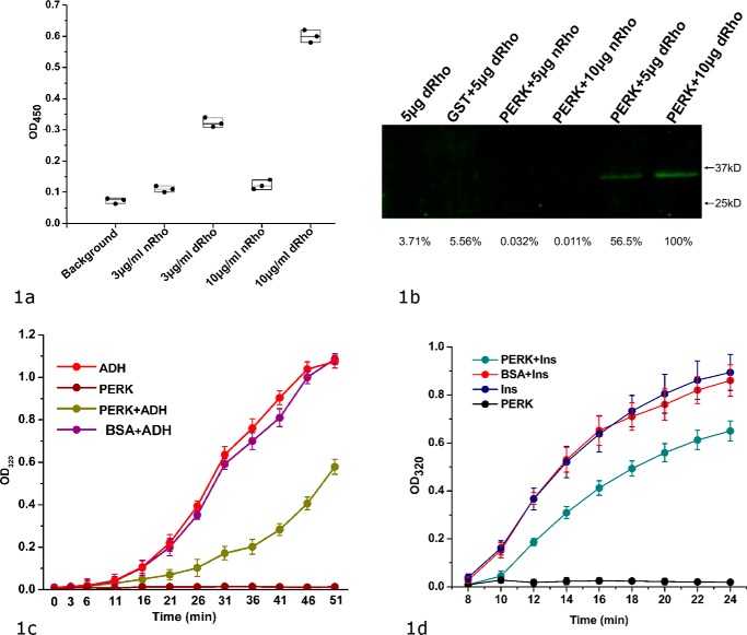
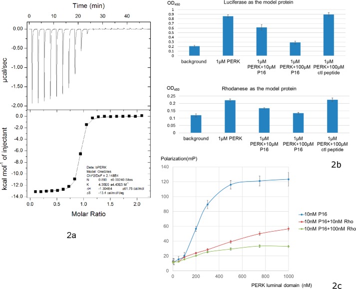
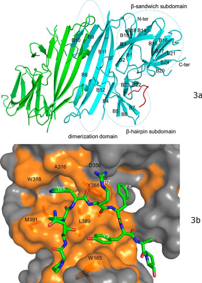
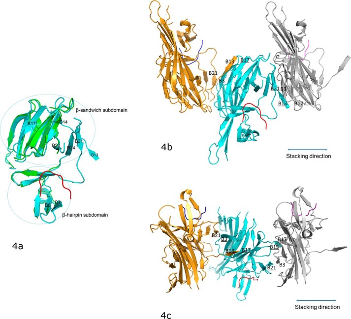
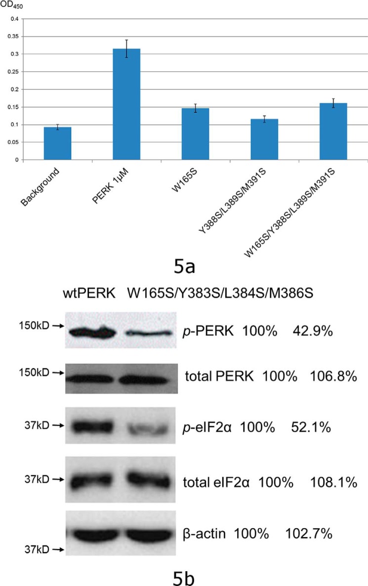
PRKR-like endoplasmic reticulum kinase (PERK) is one of the major sensor proteins that detect protein folding imbalances during endoplasmic reticulum (ER) stress. However, it remains unclear how ER stress activates PERK to initiate a downstream unfolded protein response (UPR). Here, we found that PERK's luminal domain can recognize and selectively interact with misfolded proteins but not with native proteins. Screening a phage-display library, we identified a peptide substrate, P16, of the PERK luminal domain and confirmed that P16 efficiently competes with misfolded proteins for binding this domain. To unravel the mechanism by which the PERK luminal domain interacts with misfolded proteins, we determined the crystal structure of the bovine PERK luminal domain complexed with P16 to 2.8-Å resolution. The structure revealed that PERK's luminal domain binds the peptide through a conserved hydrophobic groove. Substitutions within hydrophobic regions of the PERK luminal domain abolished the binding between PERK and misfolded proteins. We also noted that peptide binding results in major conformational changes in the PERK luminal domain that may favor PERK oligomerization. The structure of the PERK luminal domain-P16 complex suggested stacking of the luminal domain that leads to PERK oligomerization and activation via autophosphorylation after ligand binding. Collectively, our structural and biochemical results strongly support a ligand-driven model in which the PERK luminal domain interacts directly with misfolded proteins to induce PERK oligomerization and activation, resulting in ER stress signaling and the UPR.

Keywords: ER stress activation; PERK; crystal structure; crystallography; eukaryotic translation initiation factor 2α kinase 3; signaling; stress response; structural biology; unfolded protein response (UPR).


Conflict of interest statement

The authors declare that they have no conflicts of interest with the contents of this article

Figures

Figure 1.
Figure 1.
bPERK-LD can directly interact with the denatured model proteins and suppress the heat- or chemical-induced protein aggregations. a, direct binding between bovine PERK luminal domain and the denatured rhodanese shown by ELISA. The chemical denatured rhodanese (labeled as dRho) at various concentrations (0 in background, 3 and 10 μg/ml) were coated on the plate. Native rhodanese (labeled as nRho) was coated on the plate as well. The blank well was utilized as control (labeled as background). After blocking the plate by BSA, bovine PERK luminal domain at 35 μg/ml was added into the wells. After washing, the bound PERK luminal domain was detected by using anti-His tag antibody (see “Experimental procedures” for details). The OD450 readings of the individual experiments are shown in scattered dots. b, the direct binding between bovine PERK luminal domain and the denatured rhodanese shown by a pulldown assay. 100 μg of GST–bPERK-LD was mixed with 20 μl of glutathione-Sepharose 4B beads. By washing the beads with PBS buffer, different amounts of the native rhodanese (nRho) or denatured rhodanese (dRho) were applied. The beads were thoroughly washed with PBST, and then treated with the elution buffer. The eluted samples were resolved by SDS-PAGE and Western immunoblotted with antibodies against rhodanese. Naked beads with denatured rhodanese (lane 1) and purified GST (100 μg total) with denatured rhodanase (lane 2) were utilized as controls. The bands were quantified using the very right one as a reference of 100%. c, the protein aggregation suppression assay for bovine PERK luminal domain by using ADH as the model protein. 5 μm PERK LD and 10 μm ADH were utilized in this reaction. Heat-induced ADH aggregation was monitored by light scattering at OD320 at 5-min intervals. The OD320 readings are shown in vertical axis (as the percentage of the maximum value of buffer controls) and time in minutes is indicated in horizontal axis. ADH only and bPERK-LD only in PBS were used as the negative controls. 5 μm BSA mixed with 10 μm ADH was used as another control. d, protein aggregation suppression assay for the PERK luminal domain using insulin (Ins) as the model protein. Insulin (100 μm) was mixed at 25 °C with bPERK-LD (10 μm). The aggregation was induced by addition of 20 mm DTT and turbidity was monitored at 320 nm. Insulin only and bPERK-LD only were used as the negative controls. The standard derivations of three independent experiments are indicated in the bars.
Figure 2.
Figure 2.
Identification of peptide substrate P16 for bovine PERK luminal domain. a, ITC data of bovine PERK luminal domain with peptide P16. The top panel shows the heat release data for injecting the buffer containing peptide P16 in the buffer containing PERK luminal domain. Twenty injections were performed. The lower panel shows the data fitting for the released heat from the reactions with the standard model curve. b, ELISA competition assays showing the identified peptide substrate P16 can inhibit the binding capability of PERK luminal domain to the denatured model proteins luciferase (top panel) and rhodanese (lower panel) in a dose-dependent manner. 100 μl of denatured model proteins at 20 μg/ml was used to coat the plate. After washing, 100 μl of PERK luminal domain (labeled as PERK in the figure) at 1 μm was mixed with the peptide substrate P16 at 0, 10, and 100 μm, and added into the wells. The bound PERK luminal domain can be detected using anti-His tag antibody. The control 12-mer peptide GGGSGGGSGGGS (labeled as ctl peptide) showed no apparent inhibition for the binding. c, the fluorescence polarization assay to measure direct binding between PERK luminal domain and the peptide substrate P16. 10 nm 5-FAM-labeled P16 (final concentration) was added into the PERK luminal domains in 20 mm Hepes, pH 7.2, 150 mm NaCl. The concentrations of PERK luminal domain ranged from 1 to 1000 nm. After a 15-min incubation, the fluorescence polarization signals were measured. To test whether the denatured model protein rhodanese can compete with the peptide substrate P16 to bind with bovine PERK luminal domain, 10 and 100 nm denatured rhodanese were added into the reaction, respectively, and the fluorescence polarization signals were measured.
Figure 3.
Figure 3.
The crystal structure of bovine PERK luminal domain complexed with the peptide substrate P16 determined to 2.8-Å resolution. a, the PERK luminal domain homodimer structure. One of the monomers is ligand-free (in green) and the other is complexed with the peptide substrate P16 (in cyan). The peptide substrate P16 is shown in red. The N terminus and C terminus of the protein are labeled. The 24 β-strands in the complex monomer are labeled as B1–B24. The dimerization domain, β-sandwich subdomain, and β-hairpin subdomain are circled and labeled. b, the surface drawing for the PERK luminal domain complexed with the peptide substrate P16. The PERK luminal domain is shown in surface drawing and the peptide substrate P16 is shown in stick mode. The hydrophobic patches within the peptide-binding groove of the PERK luminal domain is shown in gold. The conserved residues from the peptide-binding grooves that are involved in binding the peptide substrate are labeled in black. Residues Trp-165, Tyr-388, Leu-389, and Met-391 of the PERK luminal domain are shown under the semi-transparent surface. The residues from the peptide substrate P16 that make major contacts with the PERK luminal domain are labeled in white.
Figure 4.
Figure 4.
The conformational changes of PERK luminal domain after peptide substrate binding that mediate the PERK oligomerization. a, the superimposition of the peptide-binding domains for the ligand-free monomer (in green) and the peptide-binding monomer (in cyan). The peptide substrate is in red. The β-strands where major conformational changes occur after the peptide binding are labeled. The orientation of this panel is similar to that in Fig. 3b. b, the stacking format revealed by the crystal packing for the PERK luminal domains complexed with peptide substrates. The middle PERK luminal domain complex monomer (cyan) is stacked by two neighboring complex monomers that are translated by 1 unit cell along the a axis. The stacking interactions involve two β-strand formations on either side of the middle monomer complex. On the left side, B3 and B17 (underlined) from the middle monomer form β-strands with B21 and B13 from the left monomer complex (in gold). On the right side, B13 and B21 (underlined) from the middle monomer complex form β-strands with B17 and B3 from the right monomer complex (in silver). This stacking format will allow PERK molecules to oligomerize along the stacking direction (shown in double-ended line) to both ends by any number. The orientation for the middle monomer in this panel is similar to that in panel a and Fig. 3b. c, this panel is generate by rotating b by ∼90° along the horizontal axis.
Figure 5.
Figure 5.
The structure-based mutagenesis studies. a, the mutations within the conserved hydrophobic peptide-binding groove of the PERK luminal domain comprise the abilities for the PERK luminal domain to interact with the denatured rhodanese as shown by the ELISA. The chemical denatured rhodanese at 2 μg/ml was coated on the plate. The blank well was utilized as control (labeled as background). After blocking the plate by BSA, bovine PERK luminal domain and its mutants at 1 μm (35 μg/ml) were added into the wells. After washing, the bound PERK luminal domain was detected by using anti-His tag antibody (see “Experimental procedures” for details). The OD450 readings are shown in bars. The S.D. of three independent experiments are indicated in the bars. b, the mouse PERK mutation W165S/Y383S/L384S/M386S exhibited reduced capability to sense the ER stress by use of the PERK knock-out cell line. The mouse wtPERK and PERK W165S/Y383S/L384S/M386S were transiently transfected into the PERK knock-out cell line and ER stress was induced by addition of 5 μg/ml of tunicamycin. The phosphorylated PERK (p-PERK), total PERK, phosphorylated eIF2α (p-eIF2α), and total eIF2α after ER stress were detected by Western blot. The bands were quantified using the wildtype data as a reference of 100%. The data showed that the phosphorylation levels of PERK and eIF2α were reduced by ∼50% for the PERK mutant W165S/Y383S/L384S/M386S during ER stress.

References

    1. Ron D., and Walter P. (2007) Signal integration in the endoplasmic reticulum unfolded protein response. Nat. Rev. Mol. Cell Biol. 8, 519–529 10.1038/nrm2199 - DOI - PubMed
    1. Schroder M., and Kaufman R. J. (2005) The mammalian unfolded protein response. Annu. Rev. Biochem. 74, 739–789 10.1146/annurev.biochem.73.011303.074134 - DOI - PubMed
    1. Rutkowski D. T., and Kaufman R. J. (2007) That which does not kill me makes me stronger: adapting to chronic ER stress. Trends Biochem. Sci. 32, 469–476 10.1016/j.tibs.2007.09.003 - DOI - PubMed
    1. Wek R. C., Jiang H. Y., and Anthony T. G. (2006) Coping with stress: eIF2 kinases and translational control. Biochem. Soc. Trans. 34, 7–11 10.1042/BST0340007 - DOI - PubMed
    1. Walter P., and Ron D. (2011) The unfolded protein response: from stress pathway to homeostatic regulation. Science 334, 1081–1086 - PubMed

Publication types