. 2018 Jan 31;10(426):eaao7090.
doi: 10.1126/scitranslmed.aao7090.

Zika virus-related neurotropic flaviviruses infect human placental explants and cause fetal demise in mice

Affiliations

Zika virus-related neurotropic flaviviruses infect human placental explants and cause fetal demise in mice

Derek J Platt et al. Sci Transl Med. .

Abstract

Although Zika virus (ZIKV) infection in pregnant women can cause placental damage, intrauterine growth restriction, microcephaly, and fetal demise, these disease manifestations only became apparent in the context of a large epidemic in the Americas. We hypothesized that ZIKV is not unique among arboviruses in its ability to cause congenital infection. To evaluate this, we tested the capacity of four emerging arboviruses [West Nile virus (WNV), Powassan virus (POWV), chikungunya virus (CHIKV), and Mayaro virus (MAYV)] from related (flavivirus) and unrelated (alphavirus) genera to infect the placenta and fetus in immunocompetent, wild-type mice. Although all four viruses caused placental infection, only infection with the neurotropic flaviviruses (WNV and POWV) resulted in fetal demise. WNV and POWV also replicated efficiently in second-trimester human maternal (decidua) and fetal (chorionic villi and fetal membrane) explants, whereas CHIKV and MAYV replicated less efficiently. In mice, RNA in situ hybridization and histopathological analysis revealed that WNV infected the placenta and fetal central nervous system, causing injury to the developing brain. In comparison, CHIKV and MAYV did not cause substantive placental or fetal damage despite evidence of vertical transmission. On the basis of the susceptibility of human maternal and fetal tissue explants and pathogenesis experiments in immunocompetent mice, other emerging neurotropic flaviviruses may share with ZIKV the capacity for transplacental transmission, as well as subsequent infection and injury to the developing fetus.


Conflict of interest statement

Competing interests: M.S.D. is a consultant for Inbios, Aviana, Takeda, and Sanofi-Pasteur, and is on the Scientific Advisory Boards of Moderna and OvaGene.

Figures

Fig. 1.
Fig. 1.. ZIKV-related flaviviruses cause fetal demise in mice that can be prevented by mAb treatment.
Wild-type pregnant dams were inoculated subcutaneously on E6.5 by footpad injection with 102 FFU of WNV or 103 FFU of POWV, CHIKV, MAYV, or PBS (mock) and tissues were harvested on either E13.5 or E18.5 as indicated. (A-C) qRT-PCR analysis of vRNA burden on E13.5 in the placenta (A), fetal head (B) and maternal spleen (C). (D) Fetal survival as assessed on E18.5. (E) Photograph of littermate mock- and WNV-infected fetuses at E18.5. (F) IFN-stimulated gene (ISG) mRNA expression as measured by qRT-PCR from placenta and fetal head on E13.5 after E6.5 infection with WNV, POWV, CHIKV or MAYV. (G-H) Pregnant mice were inoculated subcutaneously with WNV (102 FFU) via footpad injection on E6.5, then treated on E8.5 with a single intraperitoneal injection of 300 μg anti-WNV mAb (humanized E16) or isotype control mAb (anti-CHIKV human mAb 4N12) mAb at E8.5. Fetal heads and placentas were harvested on E13.5 for viral titer measurement by qRT-PCR (G), or on E18.5 to assess fetal survival (H). Antibody treatment experiments are pooled from 3 independent experiments with 1 to 2 pregnant female dams per experiment. vRNA burden and ISG expression data represent the mean +/− SEM for at least n = 6 tissues per group from 4 or 5 infected dams in at least 2 independent experiments. Each data point represents a biological replicate. In (A-C) and (F), data were analyzed by one-way ANOVA. In (G), data were analyzed by Mann-Whitney test (* P < 0.05; ** P < 0.01; *** P < 0.001; **** P < 0.0001). The n for survival data is indicated above each bar. Survival data were analyzed by Chi-squared test (**** P < 0.0001).
Fig. 2.
Fig. 2.. Placental histology on E18.5 after infection with WNV and CHIKV.
Wild-type pregnant mice were infected with 102 FFU of WNV or 103 FFU of CHIKV or PBS (mock) via footpad injection on E6.5, and placentas were harvested on E18.5 for histological analysis. Hematoxylin and eosin (H & E) staining of placentas was subsequently performed on mock- (A), CHIKV- (B), and WNV-infected fetuses (C). Top row = lower magnification views. Bottom two rows = higher magnification views of the decidua (D, middle row) and labyrinth (L, bottom row). SpT indicates spongiotrophoblasts within the junctional zone. (D) Placental diameter of mock-, CHIKV-, and WNV-infected placentas. Data are representative of 2 independent experiments with n = 3 tissue samples for histological analysis and n = 7 fetuses from 2 or 3 infected dams for placental diameter measurements. Scale bars = 200 μm in top row and 50 μm in bottom two rows. Data in (D) were analyzed by ANOVA test (** P < 0.01; *** P < 0.001).
Fig. 3.
Fig. 3.. Fetal brain histology on E18.5 after infection with WNV and CHIKV.
Wild-type pregnant mice were infected with 102 FFU of WNV or 103 FFU of CHIKV or PBS (mock) via footpad injection on E6.5, and fetuses were harvested on E18.5 for histological analysis. Hematoxylin and eosin (H & E) staining was performed on mock- (A), CHIKV- (B), and WNV-infected fetuses (C and D). Red arrows indicate necrosis of the brain in a severe case of WNV fetal disease. Data are representative of 2 independent experiments with n = 3 fetuses each. Scale bars = 200 μm in top rows and 100 μm in bottom rows.
Fig. 4.
Fig. 4.. WNV tissue tropism in the mouse placenta and fetus.
Wild-type pregnant mice were infected with 102 FFU of WNV or PBS (mock) via footpad injection on E6.5, and fetuses were harvested on E13.5 for analysis by RNA in situ hybridization (ISH). (A-B) Low and high-magnification images of RNA ISH staining of mock- and WNV-infected placenta on E13.5. D = decidua, JZ = junctional zone, L = labyrinth, UC = umbilical cord. Scale bars = 1 mm in (A) and 100 μm in (B). (C-G) Low and high-magnification images of RNA ISH staining of mock- and WNV-infected fetuses on E13.5. WNV RNA ISH staining in a severely infected fetuses (C), right panel and D-G), including the forebrain (D), hindbrain (E), spinal cord (F), and myocardium (G). Data are representative of 2 independent experiments with n = 3 placentas or fetuses each. Scale bars = 1 mm in (C) and 100 μm in (D-G).
Fig. 5.
Fig. 5.. Viral growth curves in human placental explants.
(A-F) Human placenta villi, decidua, and fetal membrane explants from three donors (age 21–23 weeks gestation) were inoculated with 5 × 10⁶ FFU of ZIKV, WNV, POWV (A-C), CHIKV, or MAYV (D-F), incubated at 37°C for 2.5 h, washed, and then cultured in DMEM/F12. Small aliquots of cell explant supernatants were collected at 3, 12, 24, 36, and 48 h and analyzed by focus-forming assay. Data represent the mean +/− SEM from 3 or 4 organoid explants per experiment from 3 separate donors in each condition.
Fig. 6.
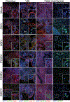
Fig. 6.. Immunofluorescence staining of virus-infected human placental explants.
Confocal micrographs of human placental tissues (chorionic villi, decidua, and fetal membrane) from four donors (age 16–23 weeks gestation) that were infected with the indicated viruses (mock (PBS), ZIKV, WNV, POWV, CHIKV, and MAYV) for 48 h. Immunofluorescence microscopy was performed for vRNA (using anti-dsRNA antibody, green) and tissue counterstained for cytokeratin-19 (a marker of epithelial-derived cells, including trophoblasts) or actin (in red, as indicated below each panel). DAPI-stained nuclei are shown in blue. White boxes denote zoomed regions shown at bottom right. Images are representative of 4 explants per experiment from 4 separate donors in each condition. Scale bar = 50 μm.

References

    1. Dick GW, Kitchen SF, Haddow AJ, Zika virus. I. Isolations and serological specificity. Transactions of the Royal Society of Tropical Medicine and Hygiene 46, 509–520 (1952). - PubMed
    1. de Oliveira WK, de França GVA, Carmo EH, Duncan BB, de Souza Kuchenbecker R, Schmidt MI, Infection-related microcephaly after the 2015 and 2016 Zika virus outbreaks in Brazil: a surveillance-based analysis. The Lancet, 390, 861–870 (2017). - PubMed
    1. Weaver SC, Emergence of Epidemic Zika Virus Transmission and Congenital Zika Syndrome: Are Recently Evolved Traits to Blame? mBio 8, pii: e02063–16 (2017). - PMC - PubMed
    1. Widman DG, Young E, Yount BL, Plante KS, Gallichotte EN, Carbaugh DL, Peck KM, Plante J, Swanstrom J, Heise MT, Lazear HM, Baric RS, A Reverse Genetics Platform That Spans the Zika Virus Family Tree. mBio 8, pii: e02014–16 (2017). - PMC - PubMed
    1. Yuan L, Huang XY, Liu ZY, Zhang F, Zhu XL, Yu JY, Ji X, Xu YP, Li G, Li C, Wang HJ, Deng YQ, Wu M, Cheng ML, Ye Q, Xie DY, Li XF, Wang X, Shi W, Hu B, Shi PY, Xu Z, Qin CF, A single mutation in the prM protein of Zika virus contributes to fetal microcephaly. Science (New York, N.Y.), 358, 933–936 (2017). - PubMed

Publication types