. 2018 Feb 13;115(7):E1550-E1559.
doi: 10.1073/pnas.1720553115. Epub 2018 Jan 31.

Platelets release pathogenic serotonin and return to circulation after immune complex-mediated sequestration

Affiliations

Platelets release pathogenic serotonin and return to circulation after immune complex-mediated sequestration

Nathalie Cloutier et al. Proc Natl Acad Sci U S A. .

Abstract

There is a growing appreciation for the contribution of platelets to immunity; however, our knowledge mostly relies on platelet functions associated with vascular injury and the prevention of bleeding. Circulating immune complexes (ICs) contribute to both chronic and acute inflammation in a multitude of clinical conditions. Herein, we scrutinized platelet responses to systemic ICs in the absence of tissue and endothelial wall injury. Platelet activation by circulating ICs through a mechanism requiring expression of platelet Fcγ receptor IIA resulted in the induction of systemic shock. IC-driven shock was dependent on release of serotonin from platelet-dense granules secondary to platelet outside-in signaling by αIIbβ3 and its ligand fibrinogen. While activated platelets sequestered in the lungs and leaky vasculature of the blood-brain barrier, platelets also sequestered in the absence of shock in mice lacking peripheral serotonin. Unexpectedly, platelets returned to the blood circulation with emptied granules and were thereby ineffective at promoting subsequent systemic shock, although they still underwent sequestration. We propose that in response to circulating ICs, platelets are a crucial mediator of the inflammatory response highly relevant to sepsis, viremia, and anaphylaxis. In addition, platelets recirculate after degranulation and sequestration, demonstrating that in adaptive immunity implicating antibody responses, activated platelets are longer lived than anticipated and may explain platelet count fluctuations in IC-driven diseases.

Keywords: Fc receptor; immune complexes; platelets; serotonin; thrombocytopenia.


Conflict of interest statement

Conflict of interest statement: J.E.I. has a financial interest in and is a founder of Platelet BioGenesis, a company that aims to produce donor-independent human platelets from human-induced pluripotent stem cells at scale. J.E.I. is an inventor on this patent. The interests of J.E.I. were reviewed and are managed by the Brigham and Women’s Hospital and Partners HealthCare in accordance with their conflict of interest policies.

Figures

Fig. 1.
Fig. 1.
Platelets are critical during the systemic response. Systemic shock (Upper) (scores defined in Materials and Methods) and temperature (Lower) were measured at the indicated time points following IC injection in FcγRIIATGN and FcγRIIAnull mice (A; n = 12); FcγRIIATGN mice preinjected with platelet-depleting antibody (PLT Ab) and isotypic control antibody (Ctrl Ab) (B; n = 8); FcγRIIATGN/β3+/+ and FcγRIIATGN/β3−/− mice (C; n = 15); bone marrow chimeric mice generated by transfer of FcγRIIATGN cells into WT (D and E; native fibrinogen), Fibγ∆5 (D), and Fibγ390-396A (E) irradiated mice (n = 6); FcγRIIATGN mice preinjected with GPIb Fab (Xia.B2) or diluent (F; n = 6); FcγRIIATGN mice pretreated with aurintricarboxylic acid (ATA) or diluent (G; n = 8); and FcγRIIATGN mice pretreated with alteplase (H; n = 4) or diluent. null, FcyRIIAnull; TGN, FcyRIIATGN. Data are mean ± SEM. **P < 0.005, ***P < 0.001, and ****P < 0.0001; repeated-measures two-way ANOVA, statistical variation between groups (AH).
Fig. 2.
Fig. 2.
Serotonin is the mediator of shock. (A) PF4 (Left) and serotonin (Right) concentrations were measured in platelet-free plasma prepared from blood of FcγRIIATGN mice collected 10 min after IC injection (n = 5). The baseline (measured in nonchallenged FcγRIIATGN mice) concentration is indicated using a dotted line. Dil, diluent. (B and C) Intravital microscopy reveals profound changes to the mouse ear vasculature in response to ICs. (B) Vascular leakage, evidenced by the presence of Evans Blue (magenta) outside blood vessels in the subendothelial matrix rich in collagen (blue), is represented using FcγRIIATGN. (Scale bar: 75 μm.) (C) Vasodilatation was measured prior to leakage (t = 8 min) in FcγRIIATGN, FcγRIIAnull, or FcγRIIATGN/Tph1−/− mice (n > 5 vessels per field in three mice per group). (DG) Systemic shock (Upper) and temperature (Lower) were measured at the indicated time points following injection of serotonin (Sero) or its Dil in FcγRIIAnull mice (D; n = 6), after IC injection in FcγRIIATGN mice treated or not treated with the SSRI fluoxetine (E; n = 10), after IC injection in FcγRIIATGN/Tph1+/+ and FcγRIIATGN/Tph1−/− mice (F; n = 9), and after injection of ICs in FcγRIIATGN mice pretreated with the 5-hydroxytryptamine receptor 2 blocker ketanserin or Dil (G; n = 5). null, FcyRIIAnull; TGN, FcyRIIATGN. Data are mean ± SEM. *P < 0.05, **P < 0.005, ***P < 0.001, and ****P < 0.0001, using an unpaired t test (A); one-way ANOVA (C); and repeated-measures two-way ANOVA, statistical variation between groups (DG).
Fig. 3.
Fig. 3.
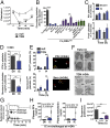
Thrombocytopenia in response to ICs. (A and B) Blood samples were collected at the indicated time points following IC injection, and CD41+ platelets in whole blood were counted using a flow cytometer. (A) Platelet count was determined in FcγRIIATGN and FcγRIIAnull mice (n = 11). (B) Percentages of circulating platelets 10 min after IC injection compared with initial platelet count were calculated in FcγRIIAnull mice (black) and the indicated three different groups of FcγRIIATGN mice. Mice were pretreated with acetylsalicylic acid (ASA) (n = 4), apyrase (n = 6), aurintricarboxylic acid (ATA) (n = 3), alteplase (n = 5), or diluent (n = 14) (all shown in blue). FcγRIIATGN mice pretreated with GPIb Fab or control (n = 4), FcγRIIATGN/β3+/+ or FcγRIIATGN/β3−/− mice (n = 10); FcγRIIATGN/Tph1+/+ mice; or FcγRIIATGN/Tph1−/− mice (n = 4) are represented (all shown in purple). Bone marrow chimeric mice generated by transfer of FcγRIIATGN cells into WT (native fibrinogen), Fibγ∆5, and Fibγ390-396A irradiated (IRRAD) mice are represented (n = 3 per group) (shown in green). (C and D) Mice were injected with diluent or IC, and blood was collected 24 h later. (C) Exposition of PS and P-selectin (P-sel) was assessed on CD41+ platelets using flow cytometry in FcγRIIATGN mice (n = 7). (D) Platelet content of PF4 (n = 6) and serotonin (n = 5) was measured by ELISA in 106 platelets retrieved 24 h after shock. Baseline (measured in nonchallenged FcγRIIATGN mice) contents are indicated using dotted lines. (E) PF4 and serotonin-negative cells were counted using immunofluorescence microscopy (n = 3 different mice). Representative image of Z-stack projections using confocal microscopy. Platelets that returned to circulation 24 h after IC injection were used for quantification. Tubulin (red) was used as a platelet marker. PF4 (green) and serotonin (green) were observed in less than 60% of platelets. Empty platelets (arrowheads) and platelets (arrows) are represented. (Scale bars: 2 μm.) As a negative control, serotonin labeling was performed on Tph1−/− platelets (Fig. S9). (F) Electron microscopy of FcγRIIATGN platelets before IC injection (Upper) and 24 h after IC injection (Lower). Empty platelets (arrowheads) and platelets (arrows) are represented. (Scale bars: 0.8 μm.) (G) Systemic shock was measured in FcγRIIATGN mice injected with ICs at t = 0 and rechallenged at t = 24 h (n = 3). (H) Plasma levels of PF4 (n = 4) and serotonin (n = 5) were determined in mice rechallenged 24 h after the first challenge with ICs. Results were compared with the level after the first challenge (dotted lines). (I) Platelets were purified from FcγRIIATGN mouse blood, fluorescently labeled, and adoptively transferred i.v. in FcγRIIATGN mice. One hour later, mice were injected with ICs, and fluorescent platelets in circulation were determined at the indicated time points following injection of ICs (n = 10). Dil, diluent; null, FcyRIIAnull; TGN; FcyRIIATGN. Data are mean ± SEM. **P < 0.005, ***P < 0.001, and ****P < 0.0001, using an unpaired t test (A, D, and H); one-way ANOVA (B, C, E, and I); and repeated-measures two-way ANOVA, statistical variation between groups (G).
Fig. 4.
Fig. 4.
Localization of sites of sequestration. (A) Mouse platelets from whole blood of FcγRIIATGN mice were labeled in vivo before IC injection. The kidneys (Kid), spleen (Spl), liver (Liv), heart (He), lungs (Lu), and brain (Br) were harvested 10 min after IC injection, and fluorescence was measured in each organ using an IVIS to determine platelet localization (n = 3). Results were compared with FcγRIIAnull mice injected with diluent, and are presented as the percentage of fluorescence in diluent-injected FcγRIIAnull mice. (B) Lungs were collected 10 min after IC injection. Thrombi (star) and neutrophils (arrows) were examined by microscopy after hematoxylin and eosin coloration. (Magnification: 400×.) (C) FcγRIIATGN mice were injected with diluent (Dil) or ICs, and lungs were collected after 10 min. The lung wet-to-dry ratio was evaluated. (D) Number of lung thrombi per square millimeter was quantified in FcγRIIAnull (n = 4), FcγRIIATGN (n = 12), FcγRIIATGN/β3−/− (n = 7), and FcγRIIATGN/Tph1−/− (n = 3) mice, as well as in FcγRIIATGN mice pretreated with GP1b Fab antibody (n = 4). (E) Mouse platelets from FcγRIIAnull and FcγRIIATGN mice were labeled in vivo. Lungs were collected 10 min after the IC trigger, and the number of fluorescent platelets was estimated (n = 4) using a standard curve designed with known numbers of fluorescent platelets spiked into nonfluorescent control lung homogenates. Percentages were obtained by comparison with platelet count obtained before the experiment and are presented as the percentage of total platelet number in the whole-mouse body. (F) ICs were labeled with DyLight-647 anti-Human IgG (fluo ICs) before injection in mice. The Kid, Spl, Liv, He, Lu, and Br were harvested 10 min later, and fluorescence was measured in each organ using an IVIS to determine IC localization (n = 3). Results are presented as the percentage of fluo determined in FcγRIIAnull mice. (G) Two-photon intravital microscopy in brain microvasculature of FcγRIIATGN/CD41-YFP mice injected with ICs. Thrombi were observed 5 min after IC injection (arrows), whereas brain vasculature leakage occurred after 10 min. (Scale bar: 50 μm.) null, FcyRIIAnull; TGN; FcyRIIATGN. Data are mean ± SEM. *P < 0.05, **P < 0.005, ***P < 0.001, and ****P < 0.0001 using one-way ANOVA (A and D) and an unpaired t test (C, E, and F).
Fig. 5.
Fig. 5.
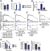
Role of the FcγRIIA/serotonin axis in acute inflammation. Endogenous antibodies directed against LPS (anti-LPS) were measured by ELISA in plasma of healthy volunteers (HV) and patients with ongoing septic shock (SS) (A; n = 11) and in FcγRIIAnull and FcγRIIATGN mice that were immunized with LPS (LPS-immune) or diluent (nonimmune) (B; n = 4). OD, optical density. (C) Systemic shock and temperature were evaluated in nonimmune (n = 4) or LPS-immune mice (n = 6) expressing or not expressing FcγRIIA. (D) Serotonin levels in plasma were determined in LPS-immune FcγRIIAnull, FcγRIIATGN, and FcγRIIATGN/β3−/− mice immediately after LPS injection (n = 5) and compared with nonimmune FcγRIIATGN mice (baseline level, indicated as a dotted line). Systemic shock and temperature were measured after LPS injection in LPS-immune FcγRIIATGN mice preinjected with platelet-depleting (PLT Ab) or control (Ctrl Ab) antibodies (E; n = 5) and in LPS-immune FcγRIIATGN/Tph1+/+ mice and FcγRIIATGN/Tph1−/− mice (F; n = 6). (G) Systemic shock was measured after LPS injection in LPS-immune FcγRIIATGN/β3+/+ mice and FcγRIIATGN/β3−/− mice (n = 4). (H) CD41+ platelets in whole blood were counted using flow cytometry at the indicated time points after injection of LPS in LPS-immune FcγRIIAnull and FcγRIIATGN mice (n = 5). (I) Platelet content in PF4 (n = 5; Left) and serotonin (n = 6; Right) were determined 48 h after LPS injection in LPS-immune FcγRIIAnull and FcγRIIATGN mice and compared with nonimmune FcγRIIATGN mouse levels (dotted lines). Dil, diluent; null, FcyRIIAnull; TGN, FcyRIIATGN. Data are mean ± SEM. **P < 0.005, ***P < 0.001, and ****P < 0.0001, using an unpaired t test (A and I); repeated-measures two-way ANOVA, statistical variation between groups (C and EG); and one-way ANOVA (B, D, and H).
Fig. 6.
Fig. 6.
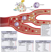
Platelets release pathogenic serotonin and return to blood circulation after IC-mediated degranulation and sequestration. Sequential events (numbered 1–6) were observed when circulating ICs encountered platelets. Platelets are abundant in blood and in humans (not in WT mice); they express FcγRIIA, a low-affinity receptor for IgG. ICs activate FcγRIIA present on platelets (1), which changes αIIbβ3 to its active conformation (2). (3) Active αIIbβ3 binds its extracellular ligand fibrinogen, which mediates outside-in signaling and granule release. In the absence of αIIbβ3, there is no granule release. It is suggested (dotted line) that serotonin engages neutrophils (4), and it is further hypothesized (dotted line) that serotonin induces vasodilatation through its action on endothelial cells (5). (6) Multiple manifestations are observed when ICs form in blood. Shock, characterized by loss of consciousness, immobility, shallow respiration, and hypothermia, strictly implicates platelets, αIIbβ3 binding to fibrinogen, and serotonin release. In the absence of neutrophils, serotonin is released but shock is abolished. It is surmised that neutrophils produce PAF in response to serotonin, which may contribute to shock downstream of serotonin release. Mediators of shock are indicated in the figure. Thrombocytopenia is due, at least in part, to platelet sequestration in certain vascular beds, notably in the lung and brain microvasculature. Sequestration implicates FcγRIIA but, in contrast to shock, occurs independent of serotonin and neutrophils, and only partially implicates β3. Thrombocytopenia is only transient, and platelets return to blood circulation with emptied granules. Microparticle release is observed in blood before return of platelets. Roles of different molecules in thrombocytopenia are indicated in the figure. Vasodilatation is implicating platelet-derived serotonin and is occurring independent of the presence of neutrophils. Thrombosis was characterized in lungs following IC administration. It implicates FcγRIIA and β3, and, in contrast to shock, it involves GPIb and not serotonin. These data support the notion that shock and thrombosis are independent events. Vascular leakage occurs independent of platelets and FcγRIIA. Molecules implicated in leakage are presented in the figure. NETosis, neutrophil extracellular traps; P-sel, P-selectin.

References

    1. Semple JW, Italiano JE, Jr, Freedman J. Platelets and the immune continuum. Nat Rev Immunol. 2011;11:264–274. - PubMed
    1. Morrell CN, Aggrey AA, Chapman LM, Modjeski KL. Emerging roles for platelets as immune and inflammatory cells. Blood. 2014;123:2759–2767. - PMC - PubMed
    1. Kapur R, Zufferey A, Boilard E, Semple JW. Nouvelle cuisine: Platelets served with inflammation. J Immunol. 2015;194:5579–5587. - PubMed
    1. Kolaczkowska E, Kubes P. Neutrophil recruitment and function in health and inflammation. Nat Rev Immunol. 2013;13:159–175. - PubMed
    1. Sreeramkumar V, et al. Neutrophils scan for activated platelets to initiate inflammation. Science. 2014;346:1234–1238. - PMC - PubMed

Publication types

MeSH terms

LinkOut - more resources