. 2018 Jan;8(1):170207.
doi: 10.1098/rsob.170207.

Identification and functional characterization of arginine vasopressin receptor 1A : atypical chemokine receptor 3 heteromers in vascular smooth muscle

Affiliations

Identification and functional characterization of arginine vasopressin receptor 1A : atypical chemokine receptor 3 heteromers in vascular smooth muscle

Lauren J Albee et al. Open Biol. 2018 Jan.

Abstract

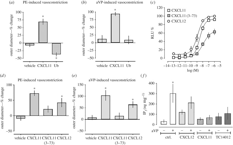
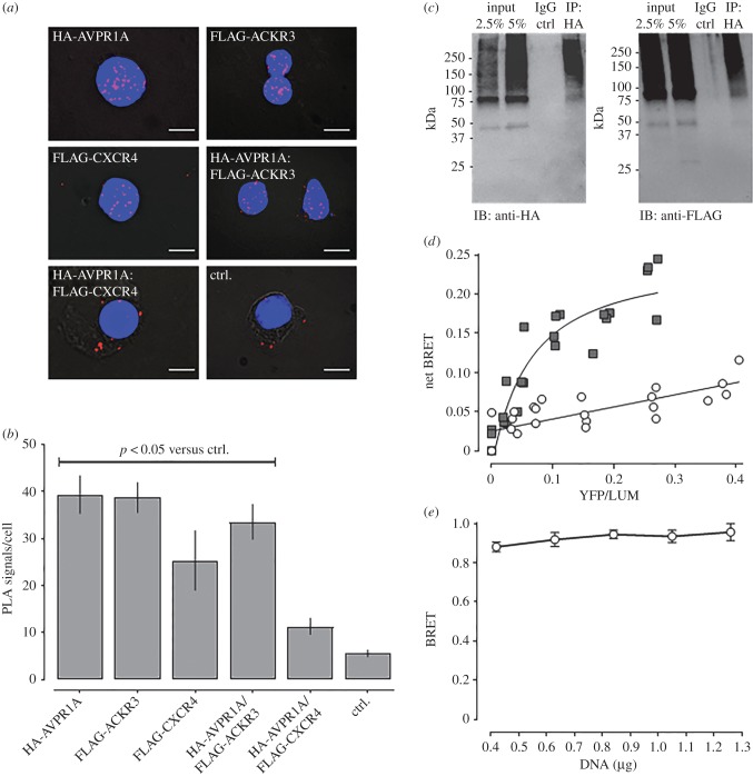
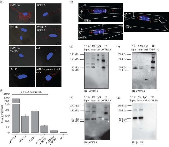
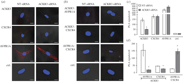
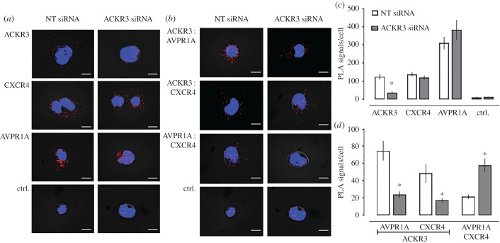
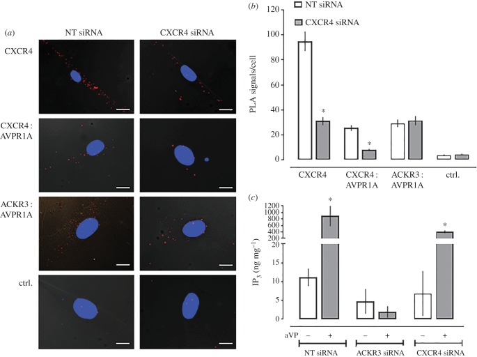
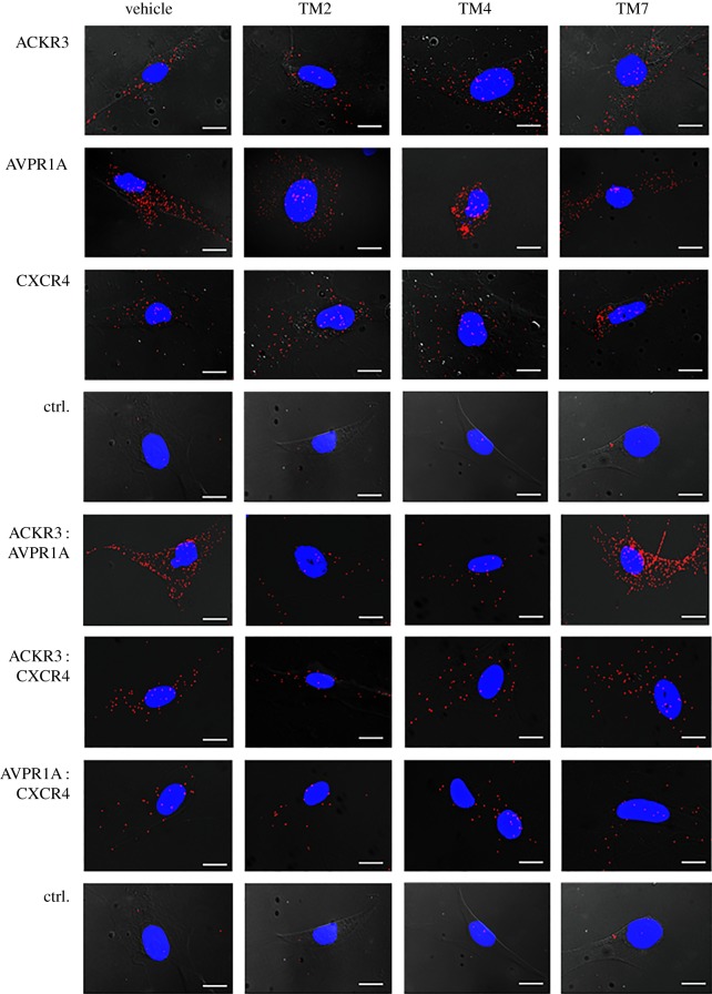
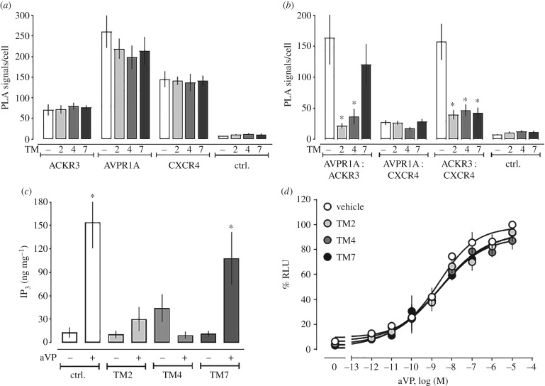
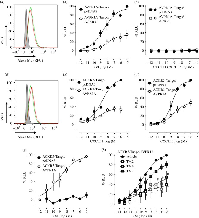
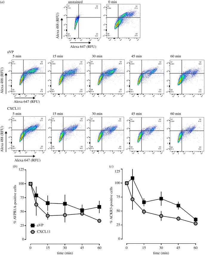
Recent observations suggest that atypical chemokine receptor (ACKR)3 and chemokine (C-X-C motif) receptor (CXCR)4 regulate human vascular smooth muscle function through hetero-oligomerization with α1-adrenoceptors. Here, we show that ACKR3 also regulates arginine vasopressin receptor (AVPR)1A. We observed that ACKR3 agonists inhibit arginine vasopressin (aVP)-induced inositol trisphosphate (IP3) production in human vascular smooth muscle cells (hVSMCs) and antagonize aVP-mediated constriction of isolated arteries. Proximity ligation assays, co-immunoprecipitation and bioluminescence resonance energy transfer experiments suggested that recombinant and endogenous ACKR3 and AVPR1A interact on the cell surface. Interference with ACKR3 : AVPR1A heteromerization using siRNA and peptide analogues of transmembrane domains of ACKR3 abolished aVP-induced IP3 production. aVP stimulation resulted in β-arrestin 2 recruitment to AVPR1A and ACKR3. While ACKR3 activation failed to cross-recruit β-arrestin 2 to AVPR1A, the presence of ACKR3 reduced the efficacy of aVP-induced β-arrestin 2 recruitment to AVPR1A. AVPR1A and ACKR3 co-internalized upon agonist stimulation in hVSMC. These data suggest that AVPR1A : ACKR3 heteromers are constitutively expressed in hVSMC, provide insights into molecular events at the heteromeric receptor complex, and offer a mechanistic basis for interactions between the innate immune and vasoactive neurohormonal systems. Our findings suggest that ACKR3 is a regulator of vascular smooth muscle function and a possible drug target in diseases associated with impaired vascular reactivity.

Keywords: CXCL11; CXCL12; arginine vasopressin; ubiquitin; vasoconstriction; vasopressor.


Conflict of interest statement

We declare we have no competing interests.

Figures

Figure 1.
Figure 1.
ACKR3 agonists antagonize aVP-mediated Gαq signalling and function in vascular smooth muscle. (a,b) Pressure myography with rat mesenteric arteries. Arteries were pressurized to 80 mmHg, preconstricted with 2 µM PE (a) or 0.5 nM aVP (b), followed by the addition of vehicle (n = 4) or 10 µM of CXCL11 (n = 6) or ubiquitin (n = 7). Outer diameter % change: % change in outer diameter after the addition of the CXCR4/ACKR3 ligands. *p < 0.05 versus vehicle. (c) β-arrestin 2 recruitment assay (PRESTO-Tango) for ACKR3. HTLA cells were treated with CXCL12, CXCL11 or CXCL11 (3–73). RLU (%): relative luminescence units in % RLU after treatment with 1 µM CXCL12 (= 100%). n = 3 independent experiments. (d) Pressure myography experiments as in (a); PE-induced vasoconstriction. All ACKR3 ligands were tested at a concentration of 10 µM. Vehicle (n = 5), CXCL11 (n = 7), CXCL11 (3–73) (n = 9) and CXCL12 (n = 14). *p < 0.05 versus vehicle. (e) Pressure myography experiments as in (b); aVP-induced vasoconstriction. All ACKR3 ligands were tested at a concentration of 10 µM. Vehicle (n = 4), CXCL11 (n = 3), CXCL11 (3–73) (n = 3) and CXCL12 (n = 3). *p < 0.05 versus vehicle. (f) hVSMCs were pretreated with either vehicle (ctrl.) or ACKR3 ligands (1 µM, 15 min) and then stimulated with 1 µM aVP for 5 min. IP3 production was measured by ELISA. n = 4. *p < 0.05 versus vehicle-treated cells.
Figure 2.
Figure 2.
Recombinant ACKR3 and AVPR1A form heteromeric complexes. (a) Typical PLA images for the detection of individual receptors and receptor–receptor interactions in HEK293T cells transfected with DNA encoding HA- or FLAG-tagged receptors. Ctrl: cells transfected with pcDNA. Images show merged PLA/DAPI signals acquired from z-stack images (n = 10; thickness 1 µm, bottom to top). Scale bars, 10 µm. (b) Quantification of PLA signals per cell as in (a). n = 3 independent experiments with n = 10 images per condition and experiment. (c) HEK293T cells expressing HA-AVPR1A and FLAG-ACKR3 were lysed (input), the lysate was immunoprecipitated (IP) with anti-HA, followed by immunoblotting (IB) to detect HA-AVPR1A (left) and FLAG-ACKR3 (right) in the IP samples. IP control: precipitate after incubation of cell lysates with IgG-coupled resin. (d,e) Intermolecular BRET assays. Cells were co-transfected with AVPR1A-hRluc plus EYFP (open circles) or ACKR3-EYFP (grey squares) at various acceptor : donor ratios (d) and with increasing amounts of AVPR1A-hRluc and ACKR3-EYFP at a constant ratio of 1 : 10 (e).
Figure 3.
Figure 3.
ACKR3 and AVPR1A form heteromeric complexes in hVSMCs. (a) Representative PLA images for the detection of individual receptors, receptor–receptor complexes and pMLC2 in hVSMC. PLA for pMLC2 was performed in non-permeabilized cells (pMLC2, bottom left) and permeabilized cells (pMLC2, permeabilized cells, bottom right). All other PLAs were performed in non-permeabilized cells. Ctrl: omission of one primary antibody. Images show merged PLA/DAPI signals acquired from z-stack images (n = 10; thickness 1 µm, bottom to top). Scale bars, 10 µm. (b) Quantification of PLA signals per cell as in (a). n = 4 independent experiments with n = 10 images per condition and experiment. (c) Three-dimensional representations of ACKR3 : AVPR1A interactions in hVSMC. Deconvolved images were generated from z-stack images (n = 20; thickness: 0.5 µm, bottom to top). Images show merged PLA/DAPI signals. (d–g) hVSMCs were lysed (=input) and AVPR1A was immunoprecipitated (IP) followed by immunoblotting (IB) to detect AVPR1A (d), CXCR4 (e), ACKR3 (f) and β2-AR (g) in the IP samples. IP control: precipitate after incubation of cell lysates with IgG-coupled resin. Images are representative of n = 4 independent experiments.
Figure 4.
Figure 4.
ACKR3 gene silencing reduces ACKR3 : AVPR1A and ACKR3 : CXCR4 heteromers and increases AVPR1A : CXCR4 interactions in hVSMCs. (a,b) Representative PLA images for the detection of individual receptors (a) and receptor–receptor interactions (b) in hVSMC after incubation with NT or ACKR3 siRNA. Ctrl: omission of one primary antibody. Images show merged PLA/4′,6-diamidino-2-phenylindole dihydrochloride (DAPI) signals acquired from z-stack images (n = 10; thickness 1 µm, bottom to top). (c,d) Quantification of PLA signals per cell for the detection of individual receptors (c) and receptor–receptor interactions (d) as in (a,b). n = 4 independent experiments with n = 10 images per condition and experiment. *p < 0.05 versus cells incubated with NT-siRNA.
Figure 5.
Figure 5.
ACKR3 gene silencing reduces ACKR3 : AVPR1A and ACKR3 : CXCR4 heteromers and increases AVPR1A : CXCR4 interactions in A7r5 cells. (a,b). Representative PLA images for the detection of individual receptors (a) and receptor–receptor interactions (b) in A7r5 cells after incubation with NT or ACKR3 siRNA. Ctrl: omission of one primary antibody. Images show merged PLA/4′,6-diamidino-2-phenylindole dihydrochloride (DAPI) signals acquired from z-stack images (n = 10; thickness 1 µm, bottom to top). (c,d) Quantification of PLA signals per cell for the detection of individual receptors (c) and receptor–receptor interactions (d) as in (a,b). n = 6 independent experiments with n = 10 images per condition and experiment. *p < 0.05 versus cells incubated with NT-siRNA.
Figure 6.
Figure 6.
ACKR3 gene silencing inhibits aVP-mediated Gαq signalling, whereas CXCR4 gene silencing does not affect ACKR3 : AVPR1A heteromerization and aVP-mediated Gαq signalling. (a) Representative PLA images for the detection of CXCR4 and receptor–receptor interactions in hVSMC after incubation with NT or CXCR4 siRNA. Ctrl: omission of one primary antibody. Images show merged PLA/4′,6-diamidino-2-phenylindole dihydrochloride (DAPI) signals acquired from z-stack images (n = 10; thickness 1 µm, bottom to top). Scale bars, 10 µm. (b) Quantification of PLA signals per cell as in (a). n = 4 independent experiments with n = 10 images per condition and experiment. *p < 0.05 versus cells incubated with NT-siRNA. (c) IP3 production of hVSMC incubated with NT, ACKR3 or CXCR4 siRNA upon stimulation with vehicle (−) or 1 µm aVP (+) for 5 min. n = 4 independent experiments. *p < 0.05 versus vehicle.
Figure 7.
Figure 7.
ACKR3-derived TM peptide analogues disrupt ACKR3 : AVPR1A and ACKR3 : CXCR4 heteromeric complexes. hVSMCs were incubated with vehicle, TM2, TM4 or TM7 (10 µM, 30 min at 37°C), washed and fixed for PLA. Typical PLA images for the detection of individual receptors or receptor–receptor complexes are shown. Ctrl: omission of one primary antibody. Images show merged PLA/4′,6-diamidino-2-phenylindole dihydrochloride (DAPI) signals acquired from z-stack images (n = 10; thickness 1 µm, bottom to top). Scale bars, 10 µm.
Figure 8.
Figure 8.
ACKR3-derived TM peptide analogues interfere with ACKR3 : AVPR1A and ACKR3 : CXCR4 heteromerization and inhibit aVP-induced Gαq signalling in hVSMC. (a,b) Quantification of PLA signals per cell as in figure 7 for individual receptors (a) and receptor–receptor interactions (b). n = 4 independent experiments with n = 10 images per condition and experiment. *p < 0.05 versus cells incubated with vehicle. (c) IP3 production was measured in hVSMC pretreated with vehicle or TM2/4/7 peptide analogues (10 µM, 30 min at 37°C) and then stimulated with either vehicle (−) or aVP (1 µM, 5 min) (+). n = 4 independent experiments. *p < 0.05 versus vehicle-treated cells. (d) β-arrestin 2 recruitment assay (PRESTO-Tango) for AVPR1A. HTLA cells expressing FLAG-AVPR1A-Tango were incubated with either vehicle or ACKR3 TM2/4/7 (10 µM, 30 min at 37°C) and then stimulated with aVP overnight. RLU (%): relative luminescence units in % RLU after treatment with 10 µM aVP (=100%). n = 3 independent experiments.
Figure 9.
Figure 9.
β-arrestin 2 recruitment at the ACKR3 : AVPR1A heteromeric complex. (a–c) HTLA cells were co-transfected with 0.75 µg of DNA for FLAG-AVPR1A-TANGO plus 0.75 µg of pcDNA3 or HA-ACKR3. (a) Measurement of FLAG-AVPR1A-Tango expression by flow cytometry. Cells were labelled with Alexa Fluor 647 anti-FLAG. Grey area: unstained cells. Red line: cells transfected with FLAG-AVPR1A-TANGO plus pcDNA. Green line: cells transfected with FLAG-AVPR1A-TANGO/HA-ACKR3. RFU: relative fluorescence units. Data are representative of n = 3 independent experiments. (b,c) β-arrestin 2 recruitment assay (PRESTO-Tango) for AVPR1A. Cells were stimulated with aVP (b), CXCL11 (c) and CXCL12 (c). Black symbols: cells transfected with FLAG-AVPR1A-TANGO/pcDNA3; white symbols: cells transfected with FLAG-AVPR1A-TANGO/HA-ACKR3. n = 3 independent experiments. (d–g) HTLA cells were co-transfected with 0.75 µg of DNA for FLAG-ACKR3-TANGO plus 0.75 µg of pcDNA3 or HA-AVPR1A. (d) Measurement of FLAG-ACKR3-Tango expression by flow cytometry. Cells were labelled with Alexa Fluor 647 anti-FLAG. Grey area: unstained cells. Red line: cells transfected with FLAG-ACKR3-TANGO plus pcDNA3. Green line: cells transfected with FLAG-ACKR3-TANGO/HA-AVPR1A. RFU: relative fluorescence units. Data are representative of n = 3 experiments. (e–g) β-arrestin 2 recruitment assay (PRESTO-Tango) for ACKR3. Cells were stimulated with CXCL11 (e), CXCL12 (f) and aVP (g). Black circles: cells transfected with FLAG-ACKR3-TANGO/pcDNA3; white circles: cells transfected with FLAG-ACKR3-TANGO/HA-AVPR1A. n = 3 independent experiments. (h) β-arrestin 2 recruitment assay (PRESTO-Tango) with HTLA cells co-expressing FLAG-AVPR1A-Tango/HA-ACKR3. Cells were treated with vehicle or TM2/4/7 peptides (10 µM, 30 min at 37°C) and then stimulated with aVP. n = 3 independent experiments. Black circle: vehicle. Open squares: TM2. Light grey squares: TM4. Dark grey squares: TM7. n = 4 independent experiments.
Figure 10.
Figure 10.
AVPR1A and ACKR3 co-internalize upon agonist stimulation in hVSMC. (a) hVSMCs were treated with 1 µM aVP or CXCL11 for up to 60 min, stained at 4°C with rabbit anti-AVPR1A/donkey anti-rabbit Alexa Fluor 647 and mouse anti-ACKR3/goat anti-mouse Alexa Fluor 488 and analysed for receptor expression via flow cytometry. RFU: relative fluorescence units. The horizontal and vertical lines show the gating thresholds for ACKR3 (Alexa 488) and AVPR1A (Alexa 647). (b) Quantification of AVPR1A-positive cells after incubation with aVP and CXCL11, as in (a). n = 3 independent experiments. (c) Quantification of ACKR3-positive cells after incubation with aVP and CXCL11, as in (a). n = 3 independent experiments.

References

    1. Gambaryan N, Perros F, Montani D, Cohen-Kaminsky S, Mazmanian M, Renaud JF, Simonneau G, Lombet A, Humbert M. 2011. Targeting of c-kit+ haematopoietic progenitor cells prevents hypoxic pulmonary hypertension. Eur. Respir. J. 37, 1392–1399. (doi:10.1183/09031936.00045710) - DOI - PubMed
    1. Yu L, Hales CA. 2011. Effect of chemokine receptor CXCR4 on hypoxia-induced pulmonary hypertension and vascular remodeling in rats. Respir. Res. 12, 21 (doi:10.1186/1465-9921-12-21) - DOI - PMC - PubMed
    1. Chu PY, Zatta A, Kiriazis H, Chin-Dusting J, Du XJ, Marshall T, Kaye DM. 2011. CXCR4 antagonism attenuates the cardiorenal consequences of mineralocorticoid excess. Circ. Heart Fail. 4, 651–658. (doi:10.1161/CIRCHEARTFAILURE.110.960831) - DOI - PubMed
    1. Sartina E, et al. 2012. Antagonism of CXCR7 attenuates chronic hypoxia-induced pulmonary hypertension. Pediatr. Res. 71, 682–688. (doi:10.1038/pr.2012.30) - DOI - PubMed
    1. Bach HH, Wong YM, LaPorte HM, Gamelli RL, Majetschak M. 2016. Pharmacological targeting of chemokine (C-X-C motif) receptor 4 in porcine polytrauma and hemorrhage models. J. Trauma Acute Care Surg. 80, 102–110. (doi:10.1097/TA.0000000000000865) - DOI - PMC - PubMed

Publication types

LinkOut - more resources