. 2018 Jan 31;8(1):1989.
doi: 10.1038/s41598-018-19755-7.

Differences in the mechanical unfolding pathways of apo- and copper-bound azurins

Affiliations

Differences in the mechanical unfolding pathways of apo- and copper-bound azurins

Anju Yadav et al. Sci Rep. .

Abstract

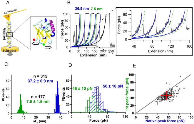
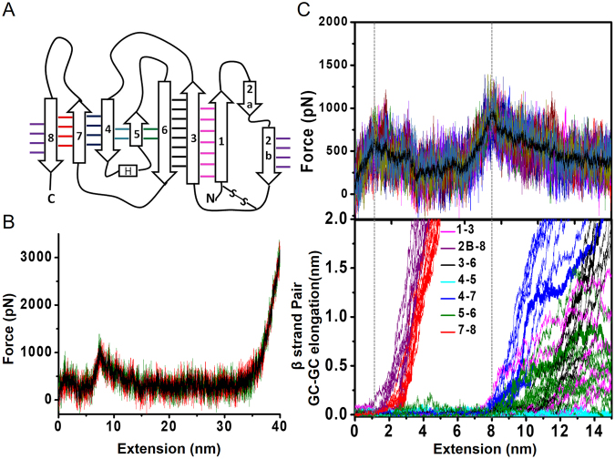
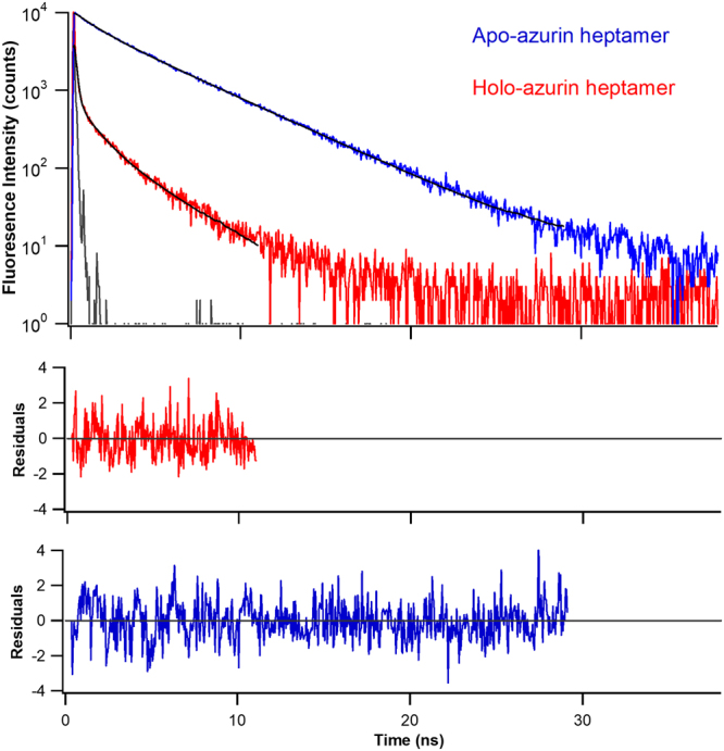
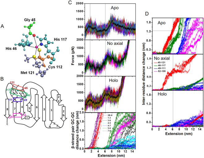
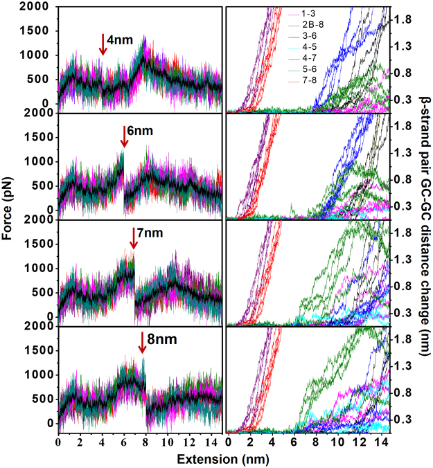
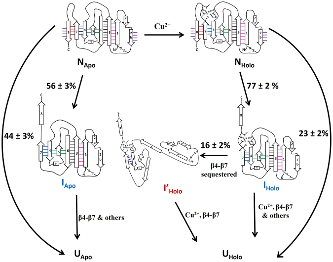
Metalloproteins carry out diverse biological functions including metal transport, electron transfer, and catalysis. At present, the influence of metal cofactors on metalloprotein stability is not well understood. Here, we report the mechanical stability and unfolding pathway of azurin, a cupredoxin family protein with β-barrel topology and type I copper-binding centre. Single-molecule force spectroscopy (SMFS) experiments reveal 2-state and 3-state unfolding pathways for apo-azurin. The intermediate in the 3-state pathway occurs at an unfolding contour length of 7.5 nm from the native state. Steered molecular dynamics (SMD) simulations show that apo-azurin unfolds via a first transition state (TS) where β2Β-β8 and β7-β8 strand pairs rupture to form the intermediate, which subsequently unfolds by the collective rupture of remaining strands. SMFS experiments on holo-azurin exhibit an additional 4-state pathway besides the 2-state and 3-state pathways. The unfolding contour length leading to the first intermediate is 6.7 nm suggesting a sequestration of ~1 nm polypeptide chain length by the copper. SMD simulations reveal atomistic details of the copper sequestration and predict a combined β4-β7 pair and copper coordination sphere rupture to create the third TS in the 4-state pathway. Our systematic studies provide detailed mechanistic insights on modulation of protein mechanical properties by metal-cofactors.


Conflict of interest statement

The authors declare that they have no competing interests.

Figures

Figure 1
Figure 1
Mechanical unfolding of apo-azurin. (A) (On left) Schematic representation of SMFS experiment on azurin polyprotein. (On right) Structure of holo-azurin (PDB code: 4AZU). (B) A representative force-versus-extension (FX) trace of apo-azurin, pulled at 400 nm/s, showing parallel unfolding pathways. Zoomed-in panel shows the trace in more detail. Worm-like chain (WLC) fits to the force peaks of native state, Napo (blue) and intermediate, Iapo (green) are shown. More FX data is given in Fig. S8. (C) Histogram of the ΔLc of the unfolding of azurin from Napo to Uapo (blue) and from Napo to Iapo (green). (D) Histogram of unfolding force of Napo (blue) and Iapo (green). Errors are SD. (E) Scatterogram of rupture forces of Iapo vs Napo for FX traces showing intermediate. Error bars are SD.
Figure 2
Figure 2
Steered molecular dynamics (SMD) simulation of apo-azurin. (A) 2D topology of apo-azurin. See Table S5 for β-strand assignment. (B) Three SMD force-versus-extension (FX) traces showing two force peaks of apo-azurin in C-terminal pulling simulations. (C) (Top) Overlay of 10 FX traces showing two TSs obtained by using different initial structures from NVT equilibrium of 100 ns. An average FX trace is also shown (solid black line). (Bottom) Geometric centres (GCs) of β-strands are calculated using the Cα atoms of the constituting residues. GC-GC distance change of adjacent β-strands as a function of unfolding extension during C-terminal pulling of apo-azurin from 10 trajectories. Data for N-terminal pulling simulations is shown in Fig. S9.
Figure 3
Figure 3
Time-resolved fluorescence intensity of heptamers of apo- and holo-azurins. Trp48 in the azurins is excited with 295 nm light. The fluorescence emission was collected at 310 nm. Instrument response function (grey solid-line) and multi-exponential fits to experimental data (black solid-lines) are shown. Residuals are shown in the bottom panels. The parameters of the fits are given in Table S1.
Figure 4
Figure 4
Mechanical unfolding of holo-azurin. (A) Representative FX trace of holo-azurin showing intermediate. Zoomed-in panel shows the trace in more detail. WLC fits are shown for the unravelling of native state, Nholo (blue) and intermediate, Iholo (green). More FX data is given in Fig. S10 in the Supplementary Material. (B) Unfolding events of holo-azurin that followed 4-state pathways: Nholo to Iholo to I’holo to Uholo. WLC fits are shown for native state, Nholo (blue), intermediate, Iholo (green), second intermediate, I’holo (red). (C) Histogram of the total change in contour length (ΔLc) of unfolding of azurin from Nholo to Uholo (blue), Nholo to Iholo (green), and Iholo to I’holo (red). Errors are SD. ΔLc histogram for Napo to Uapo (black) is also shown. (D) Histogram of unfolding force of azurin from rupture of Nholo (blue), Iholo (green), and I’holo (red). Errors are SD. (E) Scatterogram of Iholo peak force vs Nholo peak force for unfolding events showing intermediate (Iholo). Error bars are SD.
Figure 5
Figure 5
Comparison of unfolding of apo- and holo-azurins. (A) Copper coordination sphere in holo-azurin. (B) 2D topology showing inter-residue separations of the copper coordination sphere and those of the β4–β7 strand pair. (C) (Top three panels) Simulated FX traces (10 simulations) of three different forms of apo-azurin, ‘no axial’ holo-azurin, and holo-azurin. (Bottom) GC-GC distance change of adjacent β-strands as a function of C-terminal pulling of holo-azurin. All copper-ligand coordination bonds in holo-azurin and three planar equatorial copper-ligand bonds in ‘no axial’ holo-azurin were kept at a spring constant 25 kcal/mol/Å2 (see text for more details). Comparison of peak positions in FX-traces (reference dotted lines show peak positions for holo-azurin) shows that the position of the second TS peak is shifted to shorter extensions due to the presence of copper in accord with experimentally observed changes in ΔLc (Fig. 4B). Corresponding peak positions and inter-peak separations are reported in Table S6. (D) Inter-residue distance (Cα-Cα separation) change for residue pairs shown in Fig. 5B during unravelling of different forms of azurin. Thin vertical line represents the position of the second TS for holo-azurin. Thin horizontal line represents the separation of residue pair Gly45-Met121 in apo-azurin. Intersection of these two thin lines explains the larger ΔLc for the Iapo relative to that of Iholo. Note that for the ‘no axial’ model, Gly45-Met121 pair separates less relative to apo-azurin thereby producing an intermediate ΔLc between that of apo- and holo-azurins. Corresponding GC-GC distance change comparisons are provided in Fig. S13.
Figure 6
Figure 6
Controlled rupture of copper-ligand coordination sphere at different positions along the unfolding trajectory. (On left) Simulated FX traces of holo-azurin unfolding with all five copper-ligand bonds ruptured by removing harmonic constraints at different positions along the extension: 4 nm (i.e., before the second TS), 6, 7 and 8 nm (i.e., at the second TS). For each rupture condition 5 FX traces and their average (solid black line) are shown. Arrows show the position where the copper-ligands were ruptured. (On right) Corresponding GC-GC distance changes of β-strand pairs with molecular extension.
Figure 7
Figure 7
Schematic showing the unfolding mechanism of apo- and holo-azurins from their native states (Napo and Nholo) to the unfolded states (Uapo and Uholo). The flux data through the parallel mechanical pathways was obtained through SMFS experiments and the structural information on the sequence of β-strand rupture and unravelling through SMD simulations. Apo-azurin follows both 2-state and 3-state pathways, whereas holo-azurin follows 2-state, 3-state and 4-state pathways. The intermediates on the unfolding pathways of apo- and holo-azurin are different.

Similar articles

Cited by

References

    1. Dill KA, Chan HS. From Levinthal to pathways to funnels. Nat Struct Biol. 1997;4:10–9. doi: 10.1038/nsb0197-10. - DOI - PubMed
    1. Bryngelson JD, Onuchic JN, Socci ND, Wolynes PG. Funnels, pathways, and the energy landscape of protein folding: a synthesis. Proteins. 1995;21:167–95. doi: 10.1002/prot.340210302. - DOI - PubMed
    1. Stigler J, Ziegler F, Gieseke A, Gebhardt JC, Rief M. The complex folding network of single calmodulin molecules. Science. 2011;334:512–6. doi: 10.1126/science.1207598. - DOI - PubMed
    1. Rief M, Gautel M, Oesterhelt F, Fernandez JM, Gaub HE. Reversible unfolding of individual titin immunoglobulin domains by AFM. Science. 1997;276:1109–12. doi: 10.1126/science.276.5315.1109. - DOI - PubMed
    1. Aggarwal V, et al. Ligand-modulated parallel mechanical unfolding pathways of maltose-binding proteins. J Biol Chem. 2011;286:28056–65. doi: 10.1074/jbc.M111.249045. - DOI - PMC - PubMed

Publication types

LinkOut - more resources