. 2018 Jan 31;9(1):455.
doi: 10.1038/s41467-018-02858-0.

Cellular stressors contribute to the expansion of hematopoietic clones of varying leukemic potential

Affiliations

Cellular stressors contribute to the expansion of hematopoietic clones of varying leukemic potential

Terrence N Wong et al. Nat Commun. .

Abstract

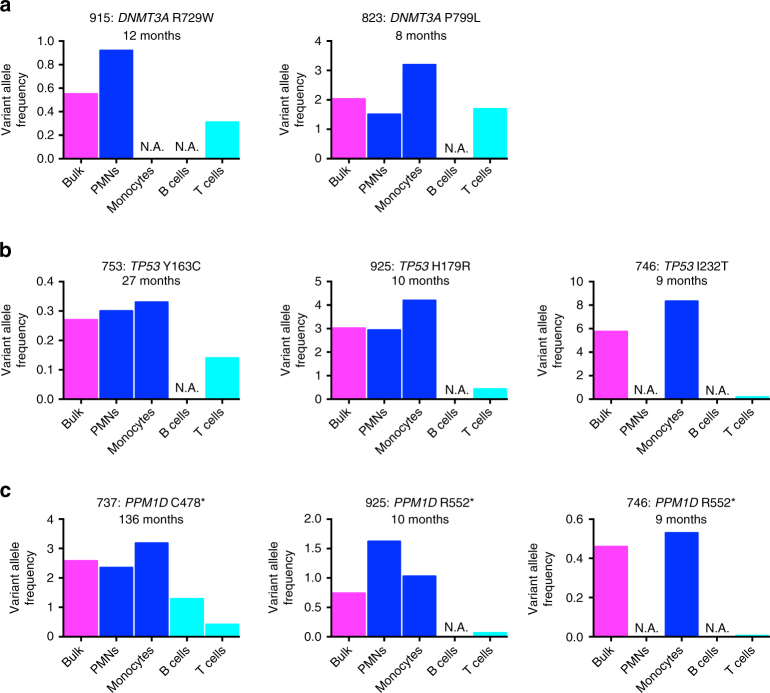
Hematopoietic clones harboring specific mutations may expand over time. However, it remains unclear how different cellular stressors influence this expansion. Here we characterize clonal hematopoiesis after two different cellular stressors: cytotoxic therapy and hematopoietic transplantation. Cytotoxic therapy results in the expansion of clones carrying mutations in DNA damage response genes, including TP53 and PPM1D. Analyses of sorted populations show that these clones are typically multilineage and myeloid-biased. Following autologous transplantation, most clones persist with stable chimerism. However, DNMT3A mutant clones often expand, while PPM1D mutant clones often decrease in size. To assess the leukemic potential of these expanded clones, we genotyped 134 t-AML/t-MDS samples. Mutations in non-TP53 DNA damage response genes are infrequent in t-AML/t-MDS despite several being commonly identified after cytotoxic therapy. These data suggest that different hematopoietic stressors promote the expansion of distinct long-lived clones, carrying specific mutations, whose leukemic potential depends partially on the mutations they harbor.


Conflict of interest statement

The authors declare no competing financial interests.

Figures

Fig. 1
Fig. 1
Influence of cytotoxic therapy on clonal expansion. a Percentage of individuals with clonal hematopoiesis ≥ 0.1% or ≥ 2.0% in three cohorts: those exposed to cytotoxic therapy (n = 81), those with malignancy but not exposed to cytotoxic therapy (n = 38), and healthy donors (n = 19). The average ages for the three groups were 55.0, 60.1, and 55.8 years, respectively. b Number of detected variants per patient. c Number of identified variants plotted against individual age for all pheresis samples. d Percentage of variants detected in pheresis samples that were transition mutations. e Percentage of individuals with at least one expanded clone harboring a variant in the specified gene. f Total number of variants identified in DNA damage response genes (i.e., TP53, PPM1D, ATM, BRCC3, SRCAP, or RAD21) grouped by whether or not the individual had previously been exposed to cytotoxic therapy. g Same as in f except for all other variants (i.e., in “non-DNA damage response genes”). h Total number of variants detected in patients with clonal hematopoiesis following cytotoxic therapy grouped by whether or not at least one variant was in a DNA damage response gene. NS not significant. *P < 0.05; **P < 0.01; ***P < 0.001. For a, d, e, significance was determined with a Fisher’s exact test. For b, significance was determined with a negative binomial regression analysis using Bonferroni-adjusted pairwise comparisons. For fh, significance was determined with a negative-binomial regression analysis. Data is represented as the mean ± the standard error of the mean
Fig. 2
Fig. 2
Expanded clones are usually multi-lineage and myeloid-biased. VAFs of the specified DNMT3A (a), TP53 (b) or PPM1D (c) mutations in bulk cells (magenta) or in sorted neutrophils (PMNs, blue), monocytes (blue), B cells (turquoise), or T cells (turquoise). N.A.: the hematopoietic population was not able to be obtained. The latency from the time of first cytotoxic therapy exposure to autologous stem cell transplant is noted
Fig. 3
Fig. 3
Influence of transplantation on clonal expansion. a Volcano plot showing the fold change in VAF following transplantation for those variants initially identified in pre-transplant pheresis samples (in blue) and those only detected after transplant (in magenta). Dotted and dashed lines respectively represent a greater than two-fold change in VAF and P < 0.05 by Fisher’s exact or χ2 tests. Triangles indicate a less than two-fold change in VAF, while circles indicate a greater than two-fold change. Variants with open circles had a greater than two-fold change but with P > 0.05. b, Summary of the changes in VAF after transplantation for all genes; changes in VAF were deemed significant if they were > 2-fold with a P value < 0.05. c, d Same as a except focusing on DNMT3A (c) or PPM1D (d) variants. e Same as b except summarizing the changes in VAF for DNMT3A and PPM1D variants. Significance for the difference in behavior after transplantation between variants in DNMT3A and PPM1D was determined with a χ2 analysis
Fig. 4
Fig. 4
Leukemogenic potential of expanded clones. a Percentage of t-AML/t-MDS cases (n = 134) harboring a mutation in the indicated gene. b Percentage of patients who after cytotoxic therapy (n=81) had an expanded clone harboring a variant in the indicated DNA damage response gene compared to the percentage of t-AML/t-MDS patients (n = 134) with a mutation in the same gene. c Same as b except that the comparison is between lymphoma patients at the time of autologous transplant (n = 69) and t-AML/t-MDS patients with a primary diagnosis of lymphoma (n = 48). For b, c significance was determined with an exact logistic regression through use of a joint test. *P < 0.05; **P < 0.01; ***P < 0.001

References

    1. Welch JS, et al. The origin and evolution of mutations in acute myeloid leukemia. Cell. 2012;150:264–278. doi: 10.1016/j.cell.2012.06.023. - DOI - PMC - PubMed
    1. Genovese G, et al. Clonal hematopoiesis and blood-cancer risk inferred from blood DNA sequence. N. Engl. J. Med. 2014;371:2477–2487. doi: 10.1056/NEJMoa1409405. - DOI - PMC - PubMed
    1. Jaiswal S, et al. Age-related clonal hematopoiesis associated with adverse outcomes. N. Engl. J. Med. 2014;371:2488–2498. doi: 10.1056/NEJMoa1408617. - DOI - PMC - PubMed
    1. Xie M, et al. Age-related mutations associated with clonal hematopoietic expansion and malignancies. Nat. Med. 2014;20:1472–1478. doi: 10.1038/nm.3733. - DOI - PMC - PubMed
    1. Steensma DP, et al. Clonal hematopoiesis of indeterminate potential and its distinction from myelodysplastic syndromes. Blood. 2015;126:9–16. doi: 10.1182/blood-2015-03-631747. - DOI - PMC - PubMed

Publication types

MeSH terms