

Reversible dysphagia secondary to guttural pouch mycosis in a gelding treated medically with voriconazole and surgically with carotid occlusion and esophagostomy
- PMID: 29386677
- PMCID: PMC5764215
Reversible dysphagia secondary to guttural pouch mycosis in a gelding treated medically with voriconazole and surgically with carotid occlusion and esophagostomy
Abstract
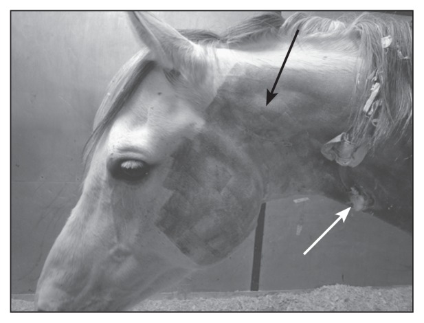
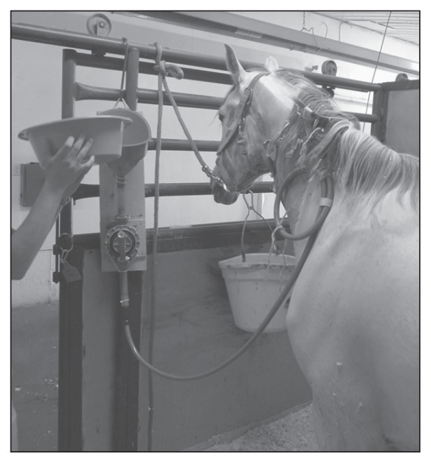
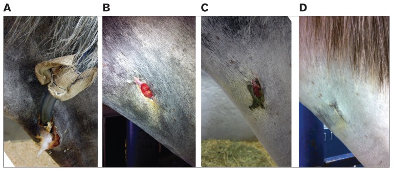
A gelding was diagnosed with dysphagia and left guttural pouch mycosis. Treatments included topical antifungal drugs, systemic voriconazole, and balloon occlusion of the internal carotid artery. Ongoing dysphagia of neurological origin necessitated extra-oral feeding through an esophagostomy tube. Complementary case management included acupuncture. Clinical remission occurred over 10 weeks.
Dysphagie réversible secondaire à une mycose de la poche gutturale chez un hongre traité médicalement avec du voriconazole et chirurgicalement par l’occlusion de la carotide et l’œsophagostomie. Un hongre a été diagnostiqué avec de la dysphagie et une mycose de la poche gutturale gauche. Les traitements ont inclus des médicaments antifongiques topiques, du voriconazole systémique et l’occlusion par ballon de l’artère carotide interne. Une dysphagie non résorbée d’origine neurologique a nécessité une alimentation extra-orale par un tube d’œsophagostomie. Une gestion du cas complémentaire a inclus l’acupuncture. Une rémission clinique s’est produite pendant 10 semaines.(Traduit par Isabelle Vallières).
Figures
Similar articles
-
Is specific antifungal therapy necessary for the treatment of guttural pouch mycosis in horses?Equine Vet J. 1995 Mar;27(2):151-2. doi: 10.1111/j.2042-3306.1995.tb03053.x. Equine Vet J. 1995. PMID: 7607150 No abstract available.
-
Successful treatment of guttural pouch mycosis with itraconazole and topical enilconazole in a horse.J Vet Intern Med. 1994 Jul-Aug;8(4):304-5. doi: 10.1111/j.1939-1676.1994.tb03239.x. J Vet Intern Med. 1994. PMID: 7983630 No abstract available.
-
Guttural pouch mycosis in horses: a retrospective study of 28 cases.Vet Rec. 2012 Dec 1;171(22):561. doi: 10.1136/vr.100700. Epub 2012 Oct 31. Vet Rec. 2012. PMID: 23118043
-
Guttural pouch diseases causing neurologic dysfunction in the horse.Vet Clin North Am Equine Pract. 2011 Dec;27(3):545-72. doi: 10.1016/j.cveq.2011.08.002. Vet Clin North Am Equine Pract. 2011. PMID: 22100044 Review.
-
The management of guttural pouch mycosis.Equine Vet J. 1989 Sep;21(5):321-4. doi: 10.1111/j.2042-3306.1989.tb02679.x. Equine Vet J. 1989. PMID: 2673759 Review. No abstract available.
References
-
- Archer RM, Knight CG, Bishop WJ. Guttural pouch mycosis in six horses in New Zealand. New Zealand Vet J. 2011;60:203–209. - PubMed
-
- Church S, Wyn-Jones G, Parks AH, Ritchie HE. Treatment of guttural pouch mycosis. Equine Vet J. 1986;18:362–365. - PubMed
-
- Cook WR. Observations on the aetiology of epistaxis and cranial nerve paralysis in the horse. Vet Rec. 1966;78:396–406. - PubMed
-
- Cook WR. The clinical features of guttural pouch mycosis in the horse. Vet Rec. 1968;83:336–345. - PubMed
-
- Dobesova O, Schwarz B, Velde K, Jahn P, Zert Z, Bezdekova B. Guttural pouch mycosis in horses: A retrospective study of 28 cases. Vet Rec. 2012;171:561. - PubMed
Publication types
MeSH terms
Substances
LinkOut - more resources
Full Text Sources
Medical