Atypical anatomy of maxillary second premolar with three roots and four canals
- PMID: 29386789
- PMCID: PMC5767836
- DOI: 10.4103/JCD.JCD_279_16
Atypical anatomy of maxillary second premolar with three roots and four canals
Abstract
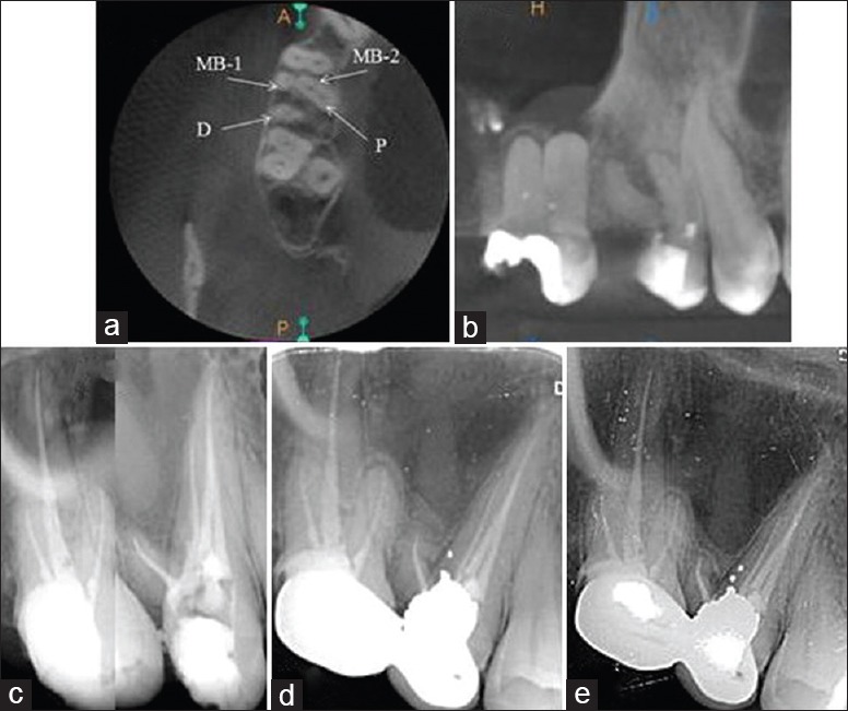
Knowledge and understanding the anatomical configuration of individual tooth play a significant role in success of endodontic treatment, in addition to through debridement and obturation of the canals. The canal anatomy of maxillary second premolar has been studied extensively, and the presence of a significant variety of multirooted canals is relatively rare in it. A 27-year-old female reported with a chief complaint of pain in her upper right posterior region for 10 days. On intraoral hard tissue examination, ill-defined access preparation was seen in maxillary right second premolar with exposed pulp. An intraoral periapical radiograph reveals radiolucency involving the pulp space and varied morphology in the same tooth. The occurrence of three roots with four canals in the maxillary second premolar is rare and not documented in the literature so far. This case report describes the nonsurgical endodontic management of such varied anatomical configuration using cone beam computed tomography as an evaluating diagnostic tool.
Keywords: Anatomical variations; cone beam computed tomography; four canals; maxillary second premolar.
Conflict of interest statement
There are no conflicts of interest.
Figures

References
-
- Cleghorn BM, Christie WH, Dong CC. The root and root canal morphology of the human mandibular first premolar: A literature review. J Endod. 2007;33:509–16. - PubMed
-
- Slowey RR. Root canal anatomy. Road map to successful endodontics. Dent Clin North Am. 1979;23:555–73. - PubMed
-
- Tian YY, Guo B, Zhang R, Yu X, Wang H, Hu T, et al. Root and canal morphology of maxillary first premolars in a Chinese subpopulation evaluated using cone-beam computed tomography. Int Endod J. 2012;45:996–1003. - PubMed
-
- Vertucci FJ. Root canal anatomy of the human permanent teeth. Oral Surg Oral Med Oral Pathol. 1984;58:589–99. - PubMed
Publication types
LinkOut - more resources
Full Text Sources
Other Literature Sources