Osteitis fibrosa cystica of mandible in hyperparathyroidism-jaw tumor syndrome: A rare presentation and review of literature
- PMID: 29386822
- PMCID: PMC5773993
- DOI: 10.4103/njms.NJMS_48_17
Osteitis fibrosa cystica of mandible in hyperparathyroidism-jaw tumor syndrome: A rare presentation and review of literature
Abstract
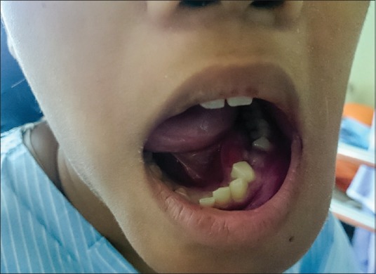
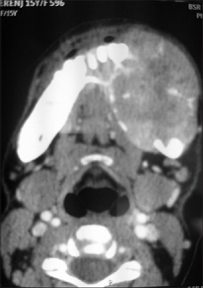
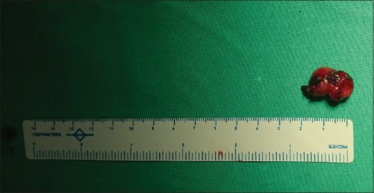
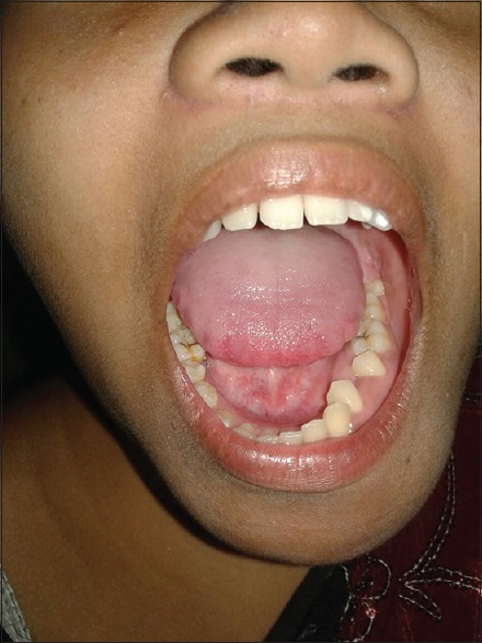
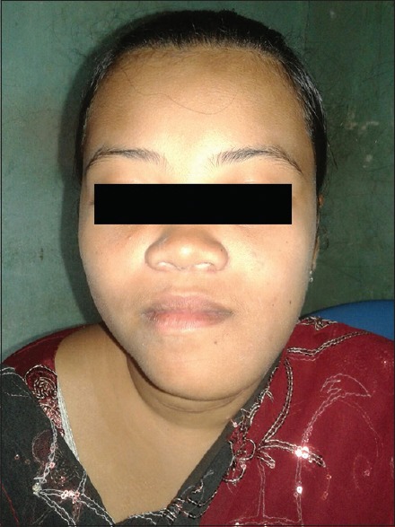
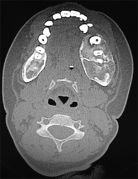
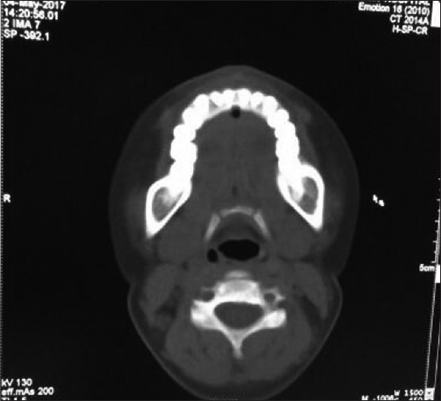
Brown's tumor, also referred as osteitis fibrosa cystica is a rare nonneoplastic diagnostically challenging consequence of hyperparathyroidism (HPT) which occurs due to increased parathormone secretions in blood, causing excessive calcium resorption from kidneys, bone resorption, and phosphaturia. Brown's tumor is a misnomer, presenting as cystic expansile lesions in bone, often misdiagnosed as neoplastic lesion or granuloma or abscess in bones. It can affect long bones, clavicle, ribs, and pelvis. According to literature, skeletal manifestations of Brown tumor is relatively rare and occurs in <2% of the cases of HPT. We present a case of a female 15-year-old patient who presented with bleeding gums and an expansile lesion in mandible whose previous investigations elsewhere suggested a malignant lesion. However, further investigations revealed it to be Brown's tumor with primary HPT which is a rare genetic disorder, known as HPT-Jaw Tumor Syndrome (HPT-JT).
Keywords: Brown tumor; hyperparathyroidism jaw tumor; mandible; parathormone.
Conflict of interest statement
There are no conflicts of interest.
Figures
Similar articles
-
Hyperparathyroidism-jaw tumour syndrome detected by aggressive generalized osteitis fibrosa cystica.Clin Cases Miner Bone Metab. 2013 Jan;10(1):65-7. doi: 10.11138/ccmbm/2013.10.1.065. Clin Cases Miner Bone Metab. 2013. PMID: 23858315 Free PMC article.
-
Tumor suppressor gene mutation in a patient with a history of hyperparathyroidism-jaw tumor syndrome and healed generalized osteitis fibrosa cystica: a case report and genetic pathophysiology review.J Oral Maxillofac Surg. 2015 Jan;73(1):194.e1-9. doi: 10.1016/j.joms.2014.09.008. Epub 2014 Sep 28. J Oral Maxillofac Surg. 2015. PMID: 25511968 Review.
-
A Large Brown Tumor of Mandible as First Manifestation of Hyperparathyroidism.J Pharm Bioallied Sci. 2024 Dec;16(Suppl 4):S3679-S3682. doi: 10.4103/jpbs.jpbs_357_24. Epub 2024 Oct 21. J Pharm Bioallied Sci. 2024. PMID: 39927001 Free PMC article.
-
Maxillofacial brown tumors in secondary hyperparathyroidism: A case report and literature review.J Res Med Sci. 2014 Nov;19(11):1099-102. J Res Med Sci. 2014. PMID: 25657758 Free PMC article.
-
Brown tumors of the jaws associated with primary or secondary hyperparathyroidism. A clinical study and review of the literature.Am J Otolaryngol. 2006 Jul-Aug;27(4):281-6. doi: 10.1016/j.amjoto.2005.11.004. Am J Otolaryngol. 2006. PMID: 16798410 Review.
Cited by
-
Osteodystrophies of jaws.J Oral Maxillofac Pathol. 2020 May-Aug;24(2):405. doi: 10.4103/jomfp.JOMFP_225_19. Epub 2020 Sep 9. J Oral Maxillofac Pathol. 2020. PMID: 33456267 Free PMC article. Review.
-
A Case of Osteitis Fibrosa Cystica of the Mandible: A Rare Presentation during Pregnancy due to CDC73 Mutation.J ASEAN Fed Endocr Soc. 2024;39(2):112-118. doi: 10.15605/jafes.039.02.17. Epub 2024 Sep 3. J ASEAN Fed Endocr Soc. 2024. PMID: 39620181 Free PMC article.
-
Brown Tumour in Chronic Kidney Disease: Revisiting an Old Disease with a New Perspective.Cancers (Basel). 2023 Aug 15;15(16):4107. doi: 10.3390/cancers15164107. Cancers (Basel). 2023. PMID: 37627135 Free PMC article. Review.
-
Parathyroid adenoma combined with a rib tumor as the primary disease: A case report.World J Clin Cases. 2020 Oct 6;8(19):4681-4687. doi: 10.12998/wjcc.v8.i19.4681. World J Clin Cases. 2020. PMID: 33083434 Free PMC article.
-
Management Modalities of Aggressive Brown Tumor of the Jaws-a Case Series.J Maxillofac Oral Surg. 2024 Jun;23(3):688-691. doi: 10.1007/s12663-024-02157-w. Epub 2024 Apr 8. J Maxillofac Oral Surg. 2024. PMID: 38911413 Free PMC article.
References
-
- Rosenberg EH, Guralnick WC. Hyperparathyroidism: A review of 220 proved cases, with special emphasis on findings in the jaw. Oral Surg Oral Med Oral Pathol. 1962;15:84–94.
-
- Ahmed R, Ahmed JM. Primary secondary, tertiary hyperparathyroidism. Otolaryngol Clin North Am. 1996;17:407–10. - PubMed
-
- Som PM, Lawson W, Cohen BA. Giant-cell lesions of the facial bones. Radiology. 1983;147:129–34. - PubMed
-
- Whitaker SB, Waldron CA. Central giant cell lesions of the jaws. A clinical, radiologic, and histopathologic study. Oral Surg Oral Med Oral Pathol. 1993;75:199–208. - PubMed
Publication types
LinkOut - more resources
Full Text Sources
Other Literature Sources