

Activity of siderophores against drug-resistant Gram-positive and Gram-negative bacteria
- PMID: 29386910
- PMCID: PMC5765970
- DOI: 10.2147/IDR.S148602
Activity of siderophores against drug-resistant Gram-positive and Gram-negative bacteria
Abstract
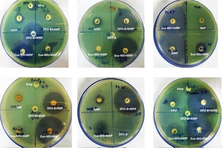
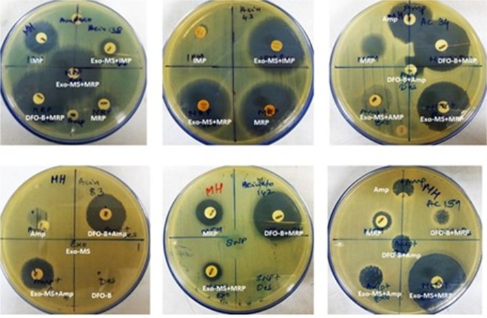
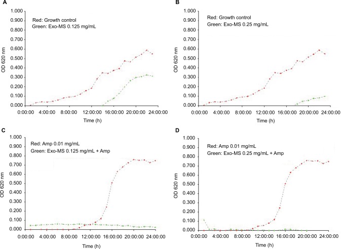
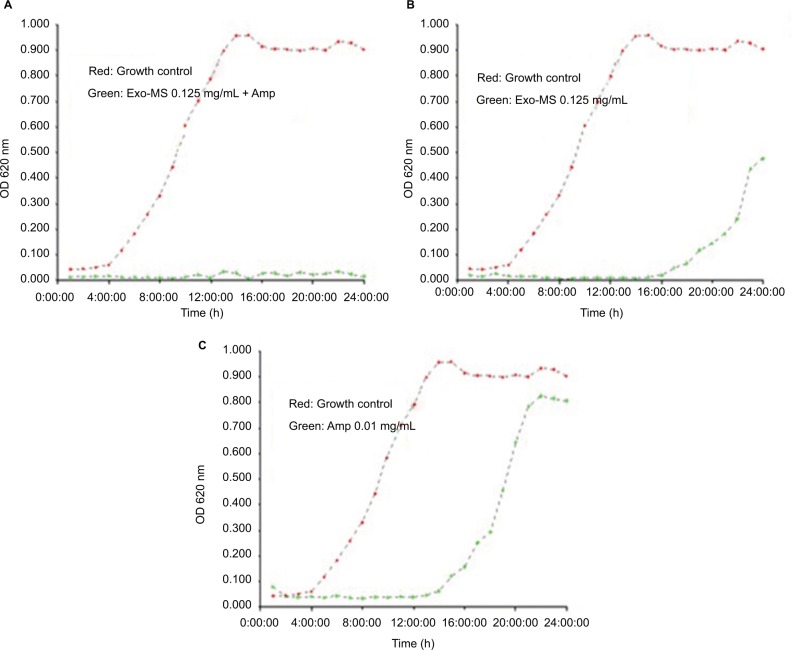
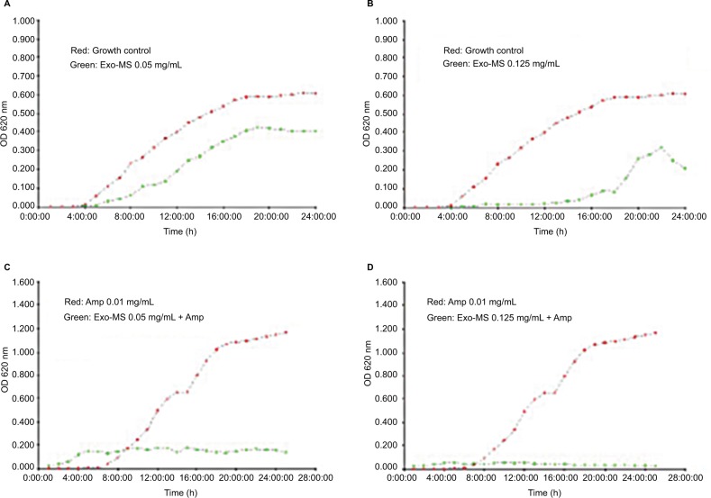
Infections by drug-resistant bacteria are life-threatening. As iron is a vital element for the growth of bacteria, iron-chelating agents (siderophores) can be used to arrest their multiplication. Exogenous siderophores - exochelin-MS and deferoxamine-B - were evaluated for their inhibitory activity against methicillin-resistant Staphylococcus aureus and metallo-β-lactamase producers - Pseudomonas aeruginosa and Acinetobacter baumannii - by disc diffusion, micro-broth dilution, and turbidimetric growth assays. The drug-resistant isolates were inhibited by the synergistic activity of siderophores and antibiotics. Minimum inhibitory concentration of exochelin-MS+ampicillin for different isolates was between 0.05 and 0.5 mg/mL. Minimum inhibitory concentration of deferoxamine-B+ampicillin was 1.0 mg/mL and greater. Iron-chelation therapy could provide a complementary approach to overcome drug resistance in pathogenic bacteria.
Keywords: deferoxamine B; exochelin MS; iron-chelation; xenosiderophores.
Conflict of interest statement
Disclosure The authors report no conflicts of interest in this work.
Figures

Similar articles
-
[Analysis of distribution and drug resistance of pathogens isolated from 541 hospitalized children with burn infection].Zhonghua Shao Shang Za Zhi. 2016 Nov 20;32(11):670-675. doi: 10.3760/cma.j.issn.1009-2587.2016.11.008. Zhonghua Shao Shang Za Zhi. 2016. PMID: 27894388 Chinese.
-
Preliminary evaluation of anti-tuberculosis potential of siderophores against drug-resistant Mycobacterium tuberculosis by mycobacteria growth indicator tube-drug sensitivity test.BMC Complement Altern Med. 2017 Mar 21;17(1):161. doi: 10.1186/s12906-017-1665-8. BMC Complement Altern Med. 2017. PMID: 28327117 Free PMC article.
-
[Analysis of distribution and drug resistance of pathogens from the wounds of 1 310 thermal burn patients].Zhonghua Shao Shang Za Zhi. 2018 Nov 20;34(11):802-808. doi: 10.3760/cma.j.issn.1009-2587.2018.11.016. Zhonghua Shao Shang Za Zhi. 2018. PMID: 30481922 Chinese.
-
Cefiderocol: A Siderophore Cephalosporin with Activity Against Carbapenem-Resistant and Multidrug-Resistant Gram-Negative Bacilli.Drugs. 2019 Feb;79(3):271-289. doi: 10.1007/s40265-019-1055-2. Drugs. 2019. PMID: 30712199 Review.
-
Treatment options for K. pneumoniae, P. aeruginosa and A. baumannii co-resistant to carbapenems, aminoglycosides, polymyxins and tigecycline: an approach based on the mechanisms of resistance to carbapenems.Infection. 2020 Dec;48(6):835-851. doi: 10.1007/s15010-020-01520-6. Epub 2020 Sep 1. Infection. 2020. PMID: 32875545 Free PMC article. Review.
Cited by
-
"Limiting access to iron decreases infection of Atlantic salmon SHK-1 cells with bacterium Piscirickettsia salmonis".BMC Vet Res. 2021 Apr 13;17(1):155. doi: 10.1186/s12917-021-02853-6. BMC Vet Res. 2021. PMID: 33849522 Free PMC article.
-
Synthesis, Photochemistry, Computational Study and Potential Application of New Styryl-Thiophene and Naphtho-Thiophene Benzylamines.Int J Mol Sci. 2022 Dec 29;24(1):610. doi: 10.3390/ijms24010610. Int J Mol Sci. 2022. PMID: 36614053 Free PMC article.
-
The application of pharmaceutical quality by design concepts to evaluate the antioxidant and antimicrobial properties of a preservative system including desferrioxamine.Daru. 2020 Dec;28(2):635-646. doi: 10.1007/s40199-020-00370-9. Epub 2020 Aug 27. Daru. 2020. PMID: 32856238 Free PMC article.
-
Fimsbactin Siderophores From a South African Marine Sponge Symbiont, Marinomonas sp. PE14-40.Microb Biotechnol. 2025 May;18(5):e70155. doi: 10.1111/1751-7915.70155. Microb Biotechnol. 2025. PMID: 40325896 Free PMC article.
-
Recent Advances in Iron Chelation and Gallium-Based Therapies for Antibiotic Resistant Bacterial Infections.Int J Mol Sci. 2021 Mar 12;22(6):2876. doi: 10.3390/ijms22062876. Int J Mol Sci. 2021. PMID: 33809032 Free PMC article. Review.
References
-
- Datta P, Gulati N, Singla N, et al. Evaluation of various methods for the detection of methicillin-resistant Staphylococcus aureus strains and susceptibility patterns. J Med Microbiol. 2011;60(Pt 11):1613–1616. - PubMed
LinkOut - more resources
Full Text Sources
Other Literature Sources
Research Materials
