. 2018 Jan 17:8:2635.
doi: 10.3389/fmicb.2017.02635. eCollection 2017.

Lauric Acid Is an Inhibitor of Clostridium difficile Growth in Vitro and Reduces Inflammation in a Mouse Infection Model

Affiliations

Lauric Acid Is an Inhibitor of Clostridium difficile Growth in Vitro and Reduces Inflammation in a Mouse Infection Model

Hsiao-Ting Yang et al. Front Microbiol. .

Abstract

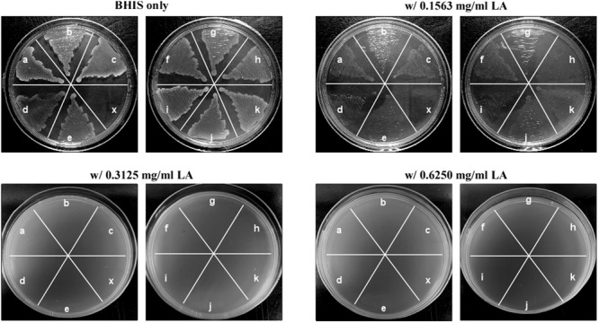
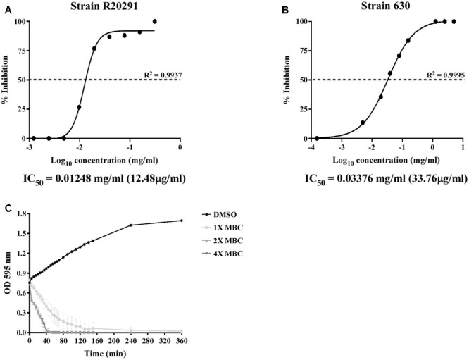
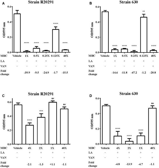
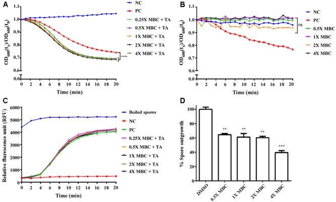
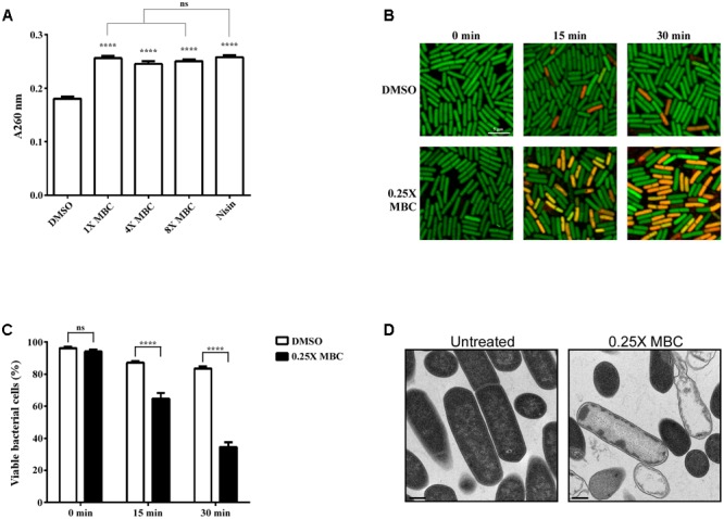
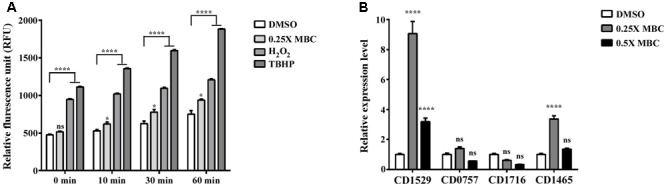
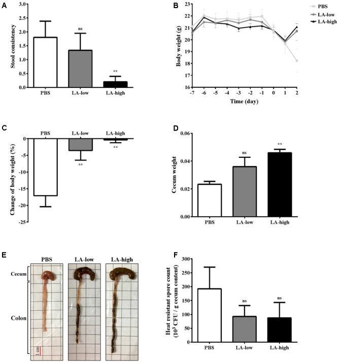
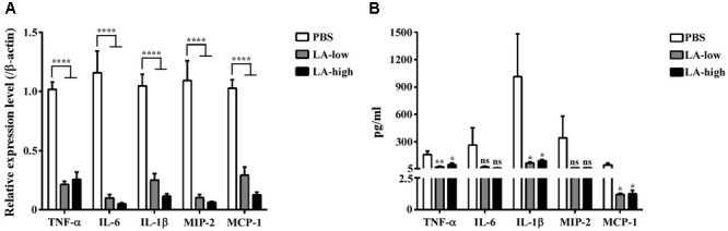
Clostridium difficile is a Gram-positive, spore-forming anaerobic human gastrointestinal pathogen. C. difficile infection (CDI) is a major health concern worldwide, with symptoms ranging from diarrhea to pseudomembranous colitis, toxic megacolon, sepsis, and death. CDI onset and progression are mostly caused by intestinal dysbiosis and exposure to C. difficile spores. Current treatment strategies include antibiotics; however, antibiotic use is often associated with high recurrence rates and an increased risk of antibiotic resistance. Medium-chain fatty acids (MCFAs) have been revealed to inhibit the growth of multiple human bacterial pathogens. Components of coconut oil, which include lauric acid, have been revealed to inhibit C. difficile growth in vitro. In this study, we demonstrated that lauric acid exhibits potent antimicrobial activities against multiple toxigenic C. difficile isolates in vitro. The inhibitory effect of lauric acid is partly due to reactive oxygen species (ROS) generation and cell membrane damage. The administration of lauric acid considerably reduced biofilm formation and preformed biofilms in a dose-dependent manner. Importantly, in a mouse infection model, lauric acid pretreatment reduced CDI symptoms and proinflammatory cytokine production. Our combined results suggest that the naturally occurring MCFA lauric acid is a novel C. difficile inhibitor and is useful in the development of an alternative or adjunctive treatment for CDI.

Keywords: Clostridium difficile; alternative therapy; lauric acid; medium-chain fatty acid; natural product.


Figures

FIGURE 1
FIGURE 1
Lauric acid is a potent inhibitor of multiple Clostridium difficile clinical isolates. Growth inhibition of multiple C. difficile isolates on BHI agar plates containing lauric acid at 0.5× (0.1563 mg/mL), 1× (0.3125 mg/mL), and 2× (0.625 mg/mL) MBC. a: TNHP 207; b: TNHP 59; c: TNHP 20; d: 630; e: R20291; f: TNHP 403; g: TNHP 82; h: TNHP 79; i: TNHP1; j: TNHP 3; k: TNHP 6; x: negative control. Results are representative of at least three independent experiments.
FIGURE 2
FIGURE 2
IC50 determination and time-dependent antibacterial kinetic curve of lauric acid against C. difficile. (A) C. difficile strains R20291 and 630 were treated with various concentrations of lauric acid for 24 h, and the OD at 600 nm was then measured. Growth inhibition was normalized to the 5% DMSO control group. (B) Log-phase grown C. difficile R20291 cells were incubated with various concentrations of lauric acid in BHI broth for up to 6 h. (C) C. difficile R20291 cells were incubated with 1×, 2×, and 4× MBC of lauric acid, and growth was monitored over time by measuring OD595. DMSO was included as a control. Results are expressed as mean of triplicate samples at least three independent experiments.
FIGURE 3
FIGURE 3
Inhibition of C. difficile biofilm by lauric acid. Various concentrations of lauric acid were added to strains R20291 (A) and 630 (B) grown in multi-well plates, and adherent biofilms were quantified by CV staining. The effect of lauric acid on preformed biofilms was measured by incubating various concentrations of lauric acid with a 24-h old biofilm formed by strains R20291 (C) and 630 (D) for an additional 24 h. The disruption of the preformed biofilm was quantified through CV staining. Vehicle: 1% DMSO only. LA: lauric acid. VAN: 20 μg/mL vancomycin (40× MBC). Lauric acid MBC: 4× = 1.25 mg/mL, 2× = 0.63 mg/mL, 1× = 0.31 mg/mL, 0.5× = 0.16 mg/mL, 0.25× = 0.08 mg/mL, 0.125× = 0.04 mg/mL. Results are the mean of triplicate samples, and one-way ANOVA was performed to assess significance. (ns, no significance; ∗∗P < 0.01, ∗∗∗P < 0.001, ∗∗∗∗P < 0.0001).
FIGURE 4
FIGURE 4
Effect of lauric acid on C. difficile spore germination. Heat-activated spores from the C. difficile strain R20291 were incubated with 10 mM sodium taurocholate plus 5% DMSO (positive control, PC), 5% DMSO only (negative control, NC), or various concentrations of lauric acid plus 10 mM sodium taurocholate and 5% DMSO. Germination was monitored by measuring absorbance at 600 nm (A,B) or DPA release (C). (D) Spore outgrowth was assayed by incubating spores in the presence of various concentrations of lauric acid for 20 min, and CFU/mL was determined by plating aliquots of spores onto BHIS agar containing TA. Results are the mean of triplicate samples, and one-way ANOVA was performed to assess significance. (ns, no significance; ∗∗P < 0.01, ∗∗∗P < 0.001, ∗∗∗∗P < 0.0001).
FIGURE 5
FIGURE 5
Lauric acid induces bacterial cell membrane damage. To measure the damaging effect of lauric acid on C. difficile cell membrane, the vegetative cells of the strain R20291 were treated with various concentrations of lauric acid for up to 30 min, and cellular material leakage was quantified by measuring absorbance at 260 nm (A). Nisin served as positive control. (B) Membrane permeability was measured by incubating cells with sublethal concentrations of lauric acid (0.25× MBC) for 15 and 30 min. Cells were then stained with SYTO9 (green) and propidium iodide (red) and imaged with confocal microscopy at 1,000× magnification. Scale bar = 5 μM. (C) Bacterial viability was quantified by counting the number of green fluorescent and red fluorescent cells from six images. (D) TEM analysis of vegetative cells treated with 0.25× MBC for 15 min compared with untreated control. Images were taken at 10,000× magnification, and scale bars = 0.5 μM. Results are the mean of three independent experiments. One-way ANOVA and two-way ANOVA was performed to assess significance for (A), and (C), respectively. ns, no significance; ∗∗∗∗P ≤ 0.0001.
FIGURE 6
FIGURE 6
Effect of lauric acid on intracellular ROS production and ROS-related genes in C. difficile. (A) C. difficile cells were treated with lauric acid for up to 60 min, and intracellular ROS was determined by staining with the general ROS indicator CM-H2DCFDA. H2O2 and TBHP served as ROS induction control. 1% DMSO – negative control. (B) The effect of lauric acid on ROS-related genes in C. difficile. C. difficile cells were treated with lauric acid or 1% DMSO for 30 min, and gene expression was then measured using real-time polymerase chain reaction. CDR20291_1529 (putative superoxide dismutase), CDR20291_0757 (putative superoxide reductase), CDR20291_1716 (putative peroxidase), and CDR20291_1465 (putative catalase). All data are presented as mean ± standard deviations, and statistical comparisons among groups were made using one-away ANOVA (p ≤ 0.05, ∗∗∗∗p ≤ 0.0001). ns, not significant. All data are representative of at least three independent experiments.
FIGURE 7
FIGURE 7
Lauric acid treatment protects mice from C. difficile infection. Various groups of mice receiving lauric acid or PBS only were treated with an antibiotic cocktail and then challenged by C. difficile for 2 days. Stool consistency (A), body weight over time (B), body weight change (C), cecum weight (D), gross views of colon and cecum (E), and heat-resistant fecal spore count 2 days post infection (F) were assessed. PBS: mice receiving PBS pre-treatment only; LA-low: mice receiving 12 mg/kg lauric acid; LA-high: mice receiving 24 mg/kg lauric acid. All data are presented as mean ± standard deviations, and statistical comparisons among groups were made using one-way ANOVA (n = 5, ∗∗P ≤ 0.01). ns, not significant. All data are representative of at least three independent experiments.
FIGURE 8
FIGURE 8
Decreased level of proinflammatory cytokines in C. difficile-infected mice receiving lauric acid. The level of various proinflammatory cytokines and chemokines in colon tissues (A) and GAL (B) from mice belonging to the PBS treatment group, LA-low (12 mg/kg) group, and LA-high (24 mg/kg) group was measured using real-time polymerase chain reactions and ELISA, respectively. All data are presented as mean ± standard deviations, and statistical comparisons among groups were made using Student’s t-test (P ≤ 0.05, ∗∗P ≤ 0.01, ∗∗∗∗P ≤ 0.0001). ns, not significant. All data are representative of at least three independent experiments.

Similar articles

Cited by

References

    1. Ababouch L., Chaibi A., Busta F. F. (1992). Inhibition of bacterial spore growth by fatty acids and their sodium salts. J. Food Prot. 55 980–984. 10.4315/0362-028x-55.12.980 - DOI - PubMed
    1. Abt M. C., McKenney P. T., Pamer E. G. (2016). Clostridium difficile colitis: pathogenesis and host defence. Nat. Rev. Microbiol. 14 609–620. 10.1038/nrmicro.2016.108 - DOI - PMC - PubMed
    1. Ananthakrishnan A. N. (2011). Clostridium difficile infection: epidemiology, risk factors and management. Nat. Rev. Gastroenterol. Hepatol. 8 17–26. 10.1038/nrgastro.2010.190 - DOI - PubMed
    1. Barbour A., Tagg J., Abou-Zied O. K., Philip K. (2016). New insights into the mode of action of the lantibiotic salivaricin B. Sci. Rep. 6:31749. 10.1038/srep31749 - DOI - PMC - PubMed
    1. Bartolotta S., Garcia C. C., Candurra N. A., Damonte E. B. (2001). Effect of fatty acids on arenavirus replication: inhibition of virus production by lauric acid. Arch. Virol. 146 777–790. - PubMed

LinkOut - more resources