. 2018 Jan;15(1):487-493.
doi: 10.3892/etm.2017.5398. Epub 2017 Oct 30.

IL-1β increases urinary corin in patients with primary proteinuric kidney diseases and in 293 cells

Affiliations

IL-1β increases urinary corin in patients with primary proteinuric kidney diseases and in 293 cells

Ci Sun et al. Exp Ther Med. 2018 Jan.

Abstract

Corin is a serine protease that is important for the regulation of blood pressure and water balance. Corin was initially discovered in the heart, however, it has also been detected in kidney cells, though its function in the kidneys is unclear. To further investigate the function of corin in the kidney, the present study analyzed the levels of corin in urine and blood samples collected from normal individuals and patients with primary proteinuric diseases. The associations between the levels of corin, and the cytokines interleukin-1β (IL-1β) and tumor necrosis factor-α (TNF-α) were then assessed. The results demonstrated that corin was detectable in the urine and plasma following an enzyme-linked immunosorbent assay; the level of corin in the urine was associated with the level of urinary β2-microglobulin (P=0.01), which was indicative of renal tubular injury. When compared with normal individuals, the levels of urinary corin in proteinuric patients were markedly increased (P=0.02), and were also associated with IL-1β (P=0.03). This correlation between corin and IL-1β was confirmed in vitro using 293 cells. As the IL-1β concentrations increased (0, 0.1, 1, 10 ng/ml), an elevation in the level of corin was observed in the culture medium (P<0.01); however, the amount of corin was not markedly altered in the cell lysate (P>0.05). In addition, when TNF-α reached 10 ng/ml, the level of corin in the medium increased significantly when compared with the control group (0 ng/ml; P=0.02), however, no significant difference in corin levels was detected in the cell lysate. The results suggest that the cytokines IL-1β and TNF-α may increase urinary corin in patients with primary proteinuric kidney diseases. Cytokines may accelerate corin shedding from the cell membrane of renal tubule epithelial cells. These findings indicate that corin may be associated with kidney inflammation and injury.

Keywords: chronic glomerulonephritis; corin protein; cytokines; interleukin-1β; tumor necrosis factor-α.


Figures

Figure 1.
Figure 1.
Corin levels in the plasma and urine. Corin could be detected in both plasma and urine samples. (A) Corin abundance in the urine of patients with proteinuric kidney diseases was higher than that in normal controls (*P=0.02). (B) Corin concentration in the plasma did not differ significantly between the patient group and the normal control group (P>0.05). (C) There was no significant correlation between plasma and urinary corin levels in normal controls (r=0.40, P>0.05). (D) There was no correlation between plasma and urinary corin levels in patients (r=0.16, P>0.05). (E) No correlation was found between urinary corin and urinary albumin (P>0.05). (F) Urinary corin was positively correlated with urinary β2-microglobulin (P=0.01). β2-MG, β2-microglobulin.
Figure 2.
Figure 2.
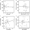
Correlation of urinary corin with CRP, eGFR, IL-1β and TNF-α. (A) No correlation was found between urinary corin and plasma CRP levels (P>0.05). (B) eGFR was not correlated with urinary corin level (P>0.05). (C) There was a positive correlation between urinary corin and plasma IL-1β levels (P=0.03). (D) No correlation existed between urinary corin and plasma TNF-α levels (P>0.05). One-way ANOVA was used for the statistical comparison among four groups. CRP, C-reactive protein; eGFR, estimated glomerular filtration rate; IL, interleukin; TNF-α, tumor necrosis factor-α.
Figure 3.
Figure 3.
Correlation between IL-1β and corin in HEK 293 cells expressing corin. (A) In the cell culture medium, corin molecules of ~180, ~160 and ~100 kDa could be detected. In the cell lysate, two bands, ~190 kDa full length corin and a ~40-kDa active form, were detected. The levels of corin in the culture medium were expressed as corin in medium/corin in lysate. As IL-1β increased, the level of corin in the cell culture medium also increased (P<0.05). (B) The amount of corin in the cell lysate did not alter significantly with different concentrations of IL-1β (P>0.05). (C) Corin abundance in the culture medium increased with the duration of IL-1β treatment (P<0.01). (D-F) Results of statistical analyses of (A), (B) and (C), respectively. **P<0.01, and ***P<0.0001. One-way ANOVA was used for the statistical comparison among four groups. IL, interleukin; HEK, human embryonic kidney.
Figure 4.
Figure 4.
Correlation between TNF-α and corin in HEK 293 cells expressing corin. (A) Correlation between TNF-α and corin in cell culture medium; the level of corin increase significantly at a TNF-α concentration of 10 ng/ml compared with 0 ng/ml (P=0.02). (B) There was no correlation between TNF-α concentration and corin level in the cell lysate. (C and D) Statistical analyses of (A) and (B), respectively. **P<0.01. One-way ANOVA was used for the statistical comparison among four groups. TNF-α, tumor necrosis factor-α; HEK, human embryonic kidney.
Figure 5.
Figure 5.
Correlation between IL-6 and corin in human embryonic kidney 293 cells expressing corin. (A) There was no correlation between IL-6 and corin level in the cell culture medium. (B) No correlation was found between IL-6 and corin level in the cell lysate. (C and D) Statistical analyses of (A) and (B), respectively. IL, interleukin.

References

    1. Hooper JD, Scarman AL, Clarke BE, Normyle JF, Antalis TM. Localization of the mosaic transmembrane serine protease corin to heart myocytes. Eur J Biochem. 2000;267:6931–6937. doi: 10.1046/j.1432-1033.2000.01806.x. - DOI - PubMed
    1. Wu Q. The serine protease corin in cardiovascular biology and disease. Front Biosci. 2007;12:4179–4190. doi: 10.2741/2379. - DOI - PubMed
    1. Gladysheva IP, Robinson BR, Houng AK, Kováts T, King SM. Corin is co-expressed with pro-ANP and localized on the cardiomyocyte surface in both zymogen and catalytically active forms. J Mol Cell Cardiol. 2008;44:131–142. doi: 10.1016/j.yjmcc.2007.10.002. - DOI - PubMed
    1. Chen S, Sen S, Young D, Wang W, Moravec CS, Wu Q. Protease corin expression and activity in failing hearts. Am J Physiol Heart Circ Physiol. 2010;299:H1687–H1692. doi: 10.1152/ajpheart.00399.2010. - DOI - PMC - PubMed
    1. Yan W, Sheng N, Seto M, Morser J, Wu Q. Corin, a mosaic transmembrane serine protease encoded by a novel cDNA from human heart. J Biol Chem. 1999;274:14926–14935. doi: 10.1074/jbc.274.21.14926. - DOI - PubMed