

Evidence of progressive tissue loss in the core of chronic MS lesions: A longitudinal DTI study
- PMID: 29387524
- PMCID: PMC5772506
- DOI: 10.1016/j.nicl.2017.12.010
Evidence of progressive tissue loss in the core of chronic MS lesions: A longitudinal DTI study
Abstract
Objective: Using diffusion tensor imaging (DTI), we examined chronic stable MS lesions, peri-lesional white matter (PLWM) and normal appearing white matter (NAWM) in patients with relapsing-remitting multiple sclerosis (RRMS) for evidence of progressive tissue destruction and evaluated whether diffusivity change is associated with conventional MRI parameters and clinical findings.
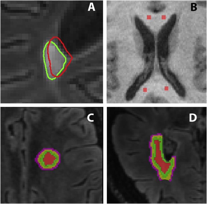
Method: Pre- and post-gadolinium T1, T2 and DTI images were acquired from 55 consecutive RRMS patients at baseline and 42.3 ± 9.7 months later. Chronic stable T2 lesions of sufficient size were identified in 43 patients (total of 134 lesions). Diffusivity parameters such as axial diffusivity (AD), radial diffusivity (RD), mean diffusivity (MD) and fractional anisotropy (FA) were compared at baseline and follow-up. MRI was also performed in 20 normal subjects of similar age and gender.
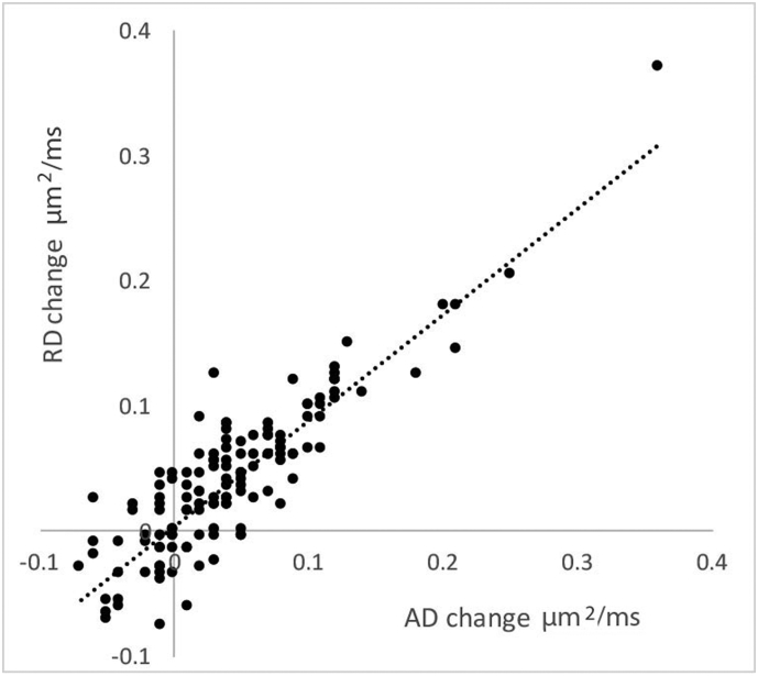
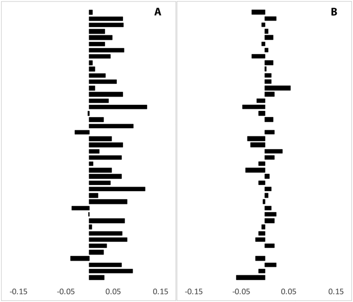
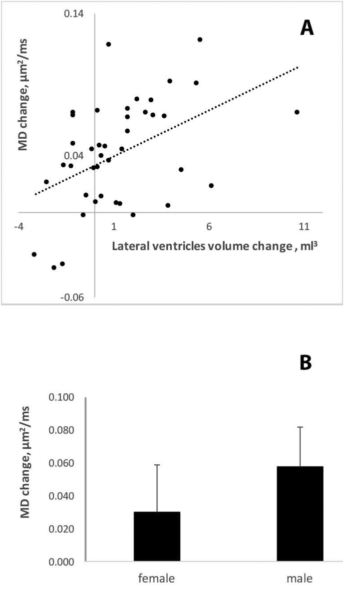
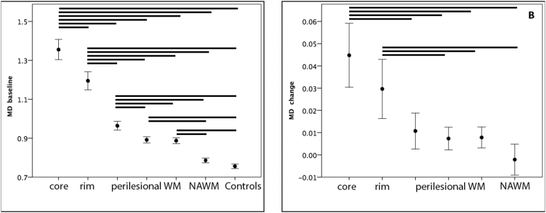
Results: Within the core of chronic MS lesions the diffusion of water molecules significantly increased over the follow-up period, while in NAWM all diffusivity indices remained stable. Since increase of AD and RD in lesional core was highly concordant, indicating isotropic nature of diffusivity change, and considering potential effect of crossing fibers on directionally-selective indices, only MD, a directionally-independent measure, was used for further analysis. The significant increase of MD in the lesion core during the follow-up period (1.29 ± 0.19 μm2/ms and 1.34 ± 0.20 μm2/ms at baseline and follow-up respectively, P < 0.0001) was independent of age or disease duration, total brain lesion volume or new lesion activity, lesion size or location and baseline tissue damage (T1 hypointensity). Change of MD in the lesion core, however, was associated with progressive brain atrophy (r = 0.47, P = 0.002). A significant gender difference was also observed: the MD change in male patients was almost twice that of female patients (0.030 ± 0.04 μm2/ms and 0.058 ± 0.03 μm2/ms in female and male respectively, P = 0.01). Sub-analysis of lesions with lesion-free surrounding revealed the largest MD increase in the lesion core, while MD progression gradually declined towards PLWM. MD in NAWM remained stable over the follow-up period.
Conclusion: The significant increase of isotropic water diffusion in the core of chronic stable MS lesions likely reflects gradual, self-sustained tissue destruction in demyelinated white matter that is more aggressive in males.
Keywords: Chronic demyelination; Diffusion; Lesions; Multiple sclerosis.
Figures
Similar articles
-
Diffusivity in the core of chronic multiple sclerosis lesions.PLoS One. 2018 Apr 25;13(4):e0194142. doi: 10.1371/journal.pone.0194142. eCollection 2018. PLoS One. 2018. PMID: 29694345 Free PMC article.
-
Diffusivity in multiple sclerosis lesions: At the cutting edge?Neuroimage Clin. 2016 Jul 5;12:219-26. doi: 10.1016/j.nicl.2016.07.003. eCollection 2016. Neuroimage Clin. 2016. PMID: 27489769 Free PMC article.
-
Microstructural alterations in different types of lesions and their perilesional white matter in relapsing-remitting multiple sclerosis based on diffusion kurtosis imaging.Mult Scler Relat Disord. 2023 Mar;71:104572. doi: 10.1016/j.msard.2023.104572. Epub 2023 Feb 19. Mult Scler Relat Disord. 2023. PMID: 36821979
-
A systematic review of microstructural abnormalities in multiple sclerosis detected with NODDI and DTI models of diffusion-weighted magnetic resonance imaging.Magn Reson Imaging. 2023 Dec;104:61-71. doi: 10.1016/j.mri.2023.09.010. Epub 2023 Sep 27. Magn Reson Imaging. 2023. PMID: 37775062
-
Brain diffusion MRI biomarkers after oncology treatments.Rep Pract Oncol Radiother. 2024 Feb 16;28(6):823-834. doi: 10.5603/rpor.98728. eCollection 2023. Rep Pract Oncol Radiother. 2024. PMID: 38515826 Free PMC article. Review.
Cited by
-
Structural and functional brain damage in women with multiple sclerosis: A mini-review of neuroimaging sex-based studies.Front Neurol. 2022 Dec 22;13:1057446. doi: 10.3389/fneur.2022.1057446. eCollection 2022. Front Neurol. 2022. PMID: 36619939 Free PMC article. Review.
-
Differentiation of hemispheric white matter lesions in migraine and multiple sclerosis with similar radiological features using advanced MRI.Front Neurosci. 2024 May 9;18:1384073. doi: 10.3389/fnins.2024.1384073. eCollection 2024. Front Neurosci. 2024. PMID: 38784095 Free PMC article.
-
circRNAs as Epigenetic Regulators of Integrity in Blood-Brain Barrier Architecture: Mechanisms and Therapeutic Strategies in Multiple Sclerosis.Cells. 2024 Aug 6;13(16):1316. doi: 10.3390/cells13161316. Cells. 2024. PMID: 39195206 Free PMC article. Review.
-
Stability of longitudinal DTI metrics in MS with treatment of injectables, fingolimod and dimethyl fumarate.Neuroradiol J. 2023 Aug;36(4):388-396. doi: 10.1177/19714009221140511. Epub 2022 Nov 17. Neuroradiol J. 2023. PMID: 36395524 Free PMC article.
-
Lesion activity and chronic demyelination are the major determinants of brain atrophy in MS.Neurol Neuroimmunol Neuroinflamm. 2019 Jul 16;6(5):e593. doi: 10.1212/NXI.0000000000000593. Print 2019 Sep. Neurol Neuroimmunol Neuroinflamm. 2019. PMID: 31454773 Free PMC article.
References
-
- Agosta F., Absinta M., Sormani M.P. In vivo assessment of cervical cord damage in MS patients: a longitudinal diffusion tensor MRI study. Brain. 2007;130:2211–2219. - PubMed
-
- Bammer R., Augustin M., Strasser-Fuchs S. Magnetic resonance diffusion tensor imaging for characterizing diffuse and focal white matter abnormalities in multiple sclerosis. Magn. Reson. Med. 2000;44(4):583–591. - PubMed
-
- Barkhof F., Bruck W., De Groot C.J.A. Remyelinated lesions in multiple sclerosis: magnetic resonance image appearance. Arch. Neurol. 2003;60:1073–1081. - PubMed
-
- Bermel R.A., Bakshi R. The measurement and clinical relevance of brain atrophy in multiple sclerosis. Lancet Neurol. 2006;5(2):158–170. - PubMed
-
- Bruck W. Inflammatory demyelination is not central to the pathogenesis of multiple sclerosis. J. Neurol. 2005;252(Suppl. 5):V/10–V/15. - PubMed
MeSH terms
LinkOut - more resources
Full Text Sources
Other Literature Sources
Medical
Miscellaneous
