

Cortical folding alterations in fetuses with isolated non-severe ventriculomegaly
- PMID: 29387528
- PMCID: PMC5790022
- DOI: 10.1016/j.nicl.2018.01.006
Cortical folding alterations in fetuses with isolated non-severe ventriculomegaly
Abstract
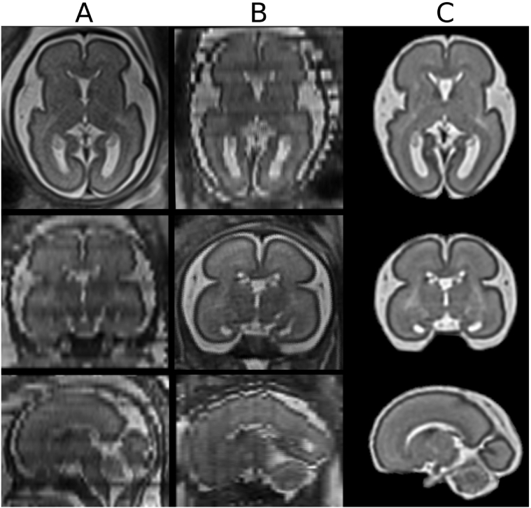
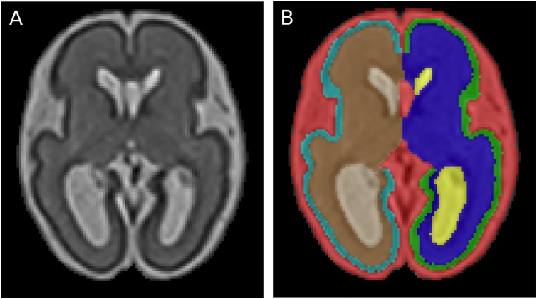
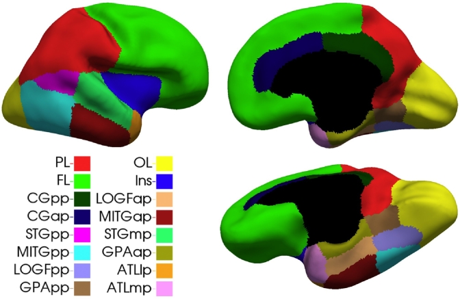
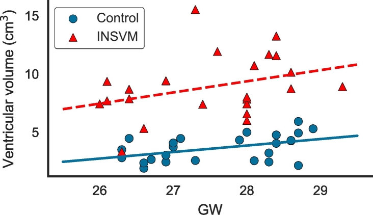
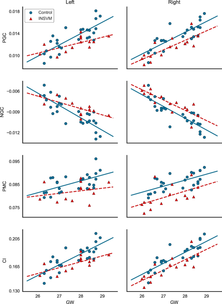
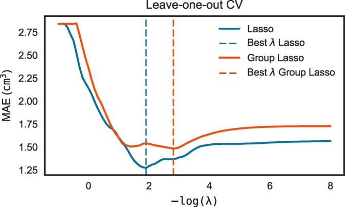
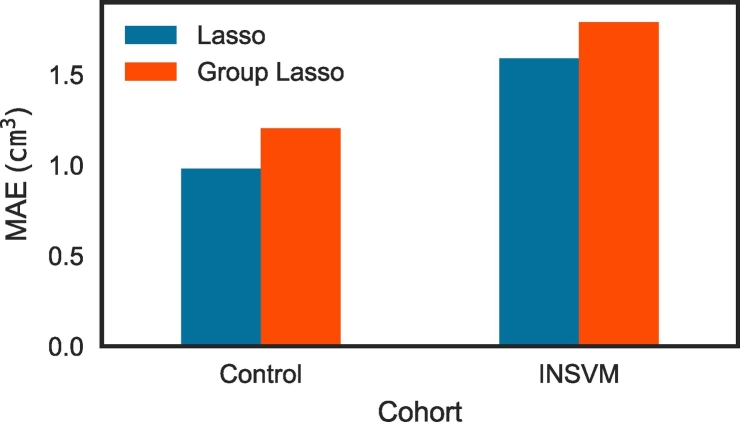
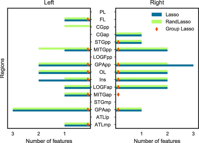
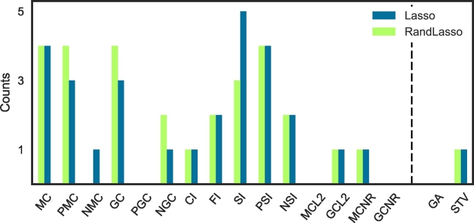
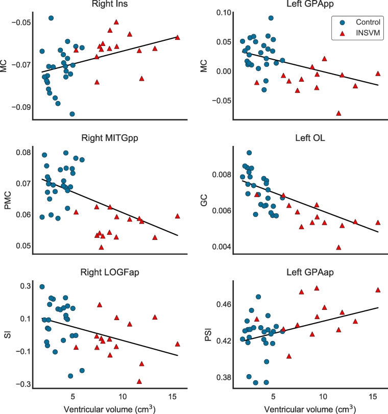
Neuroimaging of brain diseases plays a crucial role in understanding brain abnormalities and early diagnosis. Of great importance is the study of brain abnormalities in utero and the assessment of deviations in case of maldevelopment. In this work, brain magnetic resonance images from 23 isolated non-severe ventriculomegaly (INSVM) fetuses and 25 healthy controls between 26 and 29 gestational weeks were used to identify INSVM-related cortical folding deviations from normative development. Since these alterations may reflect abnormal neurodevelopment, our working hypothesis is that markers of cortical folding can provide cues to improve the prediction of later neurodevelopmental problems in INSVM subjects. We analyzed the relationship of ventricular enlargement with cortical folding alterations in a regional basis using several curvature-based measures describing the folding of each cortical region. Statistical analysis (global and hemispheric) and sparse linear regression approaches were then used to find the cortical regions whose folding is associated with ventricular dilation. Results from both approaches were in great accordance, showing a significant cortical folding decrease in the insula, posterior part of the temporal lobe and occipital lobe. Moreover, compared to the global analysis, stronger ipsilateral associations of ventricular enlargement with reduced cortical folding were encountered by the hemispheric analysis. Our findings confirm and extend previous studies by identifying various cortical regions and emphasizing ipsilateral effects of ventricular enlargement in altered folding. This suggests that INSVM is an indicator of altered cortical development, and moreover, cortical regions with reduced folding constitute potential prognostic biomarkers to be used in follow-up studies to decipher the outcome of INSVM fetuses.
Keywords: Cortical folding; Fetal brain; Lasso; MRI; Statistical analysis; Ventriculomegaly.
Figures
References
-
- Baffero G.M., Crovetto F., Fabietti I., Boito S., Fogliani R., Fumagalli M., Triulzi F., Mosca F., Fedele L., Persico N. Prenatal ultrasound predictors of postnatal major cerebral abnormalities in fetuses with apparently isolated mild ventriculomegaly. Prenat. Diagn. 2015;35:783–788. - PubMed
-
- Ball J.D., Abuhamad A.Z., Mason J.L., Burket J., Katz E., Deutsch S.I. Clinical outcomes of mild isolated cerebral ventriculomegaly in the presence of other neurodevelopmental risk factors. J. Ultrasound Med. 2013;32:1933–1938. - PubMed
-
- Batchelor P.G., Smith A.D.C., Hill D.L.G., Hawkes D.J., Cox T.C.S., Dean A.F. Measures of folding applied to the development of the human fetal brain. IEEE Trans. Med. Imaging. 2002;21:953–965. - PubMed
Publication types
MeSH terms
LinkOut - more resources
Full Text Sources
Other Literature Sources
Medical
