

Connectivity derived thalamic segmentation in deep brain stimulation for tremor
- PMID: 29387530
- PMCID: PMC5790021
- DOI: 10.1016/j.nicl.2018.01.008
Connectivity derived thalamic segmentation in deep brain stimulation for tremor
Abstract
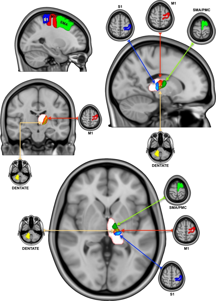
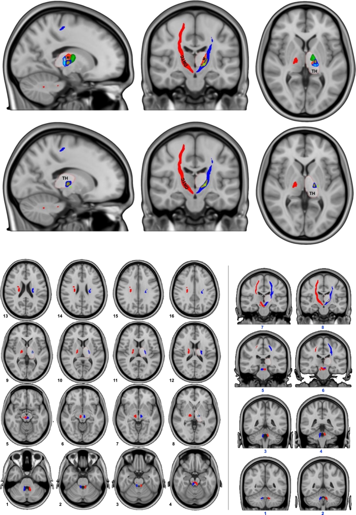
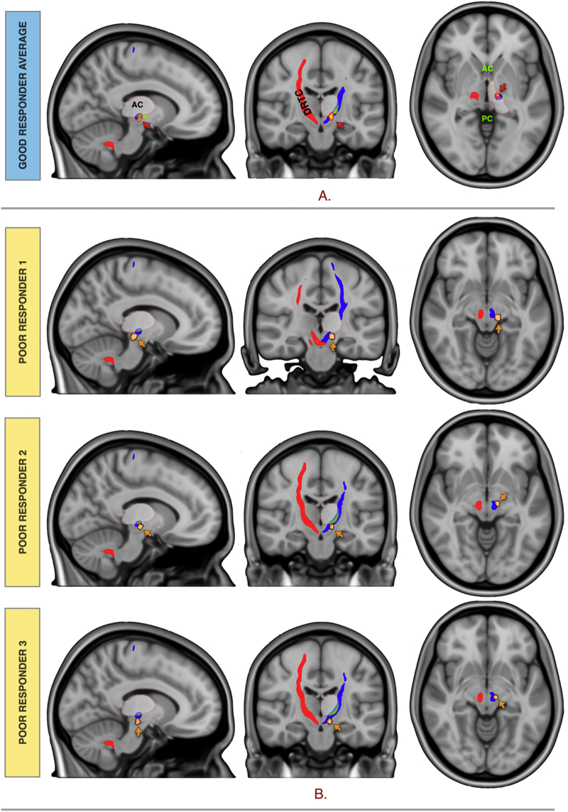
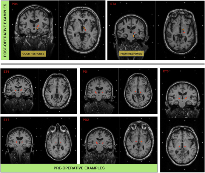
The ventral intermediate nucleus (VIM) of the thalamus is an established surgical target for stereotactic ablation and deep brain stimulation (DBS) in the treatment of tremor in Parkinson's disease (PD) and essential tremor (ET). It is centrally placed on a cerebello-thalamo-cortical network connecting the primary motor cortex, to the dentate nucleus of the contralateral cerebellum through the dentato-rubro-thalamic tract (DRT). The VIM is not readily visible on conventional MR imaging, so identifying the surgical target traditionally involved indirect targeting that relies on atlas-defined coordinates. Unfortunately, this approach does not fully account for individual variability and requires surgery to be performed with the patient awake to allow for intraoperative targeting confirmation. The aim of this study is to identify the VIM and the DRT using probabilistic tractography in patients that will undergo thalamic DBS for tremor. Four male patients with tremor dominant PD and five patients (three female) with ET underwent high angular resolution diffusion imaging (HARDI) (128 diffusion directions, 1.5 mm isotropic voxels and b value = 1500) preoperatively. Patients received VIM-DBS using an MR image guided and MR image verified approach with indirect targeting. Postoperatively, using parallel Graphical Processing Unit (GPU) processing, thalamic areas with the highest diffusion connectivity to the primary motor area (M1), supplementary motor area (SMA), primary sensory area (S1) and contralateral dentate nucleus were identified. Additionally, volume of tissue activation (VTA) corresponding to active DBS contacts were modelled. Response to treatment was defined as 40% reduction in the total Fahn-Tolosa-Martin Tremor Rating Score (FTMTRS) with DBS-ON, one year from surgery. Three out of nine patients had a suboptimal, long-term response to treatment. The segmented thalamic areas corresponded well to anatomically known counterparts in the ventrolateral (VL) and ventroposterior (VP) thalamus. The dentate-thalamic area, lay within the M1-thalamic area in a ventral and lateral location. Streamlines corresponding to the DRT connected M1 to the contralateral dentate nucleus via the dentate-thalamic area, clearly crossing the midline in the mesencephalon. Good response was seen when the active contact VTA was in the thalamic area with highest connectivity to the contralateral dentate nucleus. Non-responders had active contact VTAs outside the dentate-thalamic area. We conclude that probabilistic tractography techniques can be used to segment the VL and VP thalamus based on cortical and cerebellar connectivity. The thalamic area, best representing the VIM, is connected to the contralateral dentate cerebellar nucleus. Connectivity based segmentation of the VIM can be achieved in individual patients in a clinically feasible timescale, using HARDI and high performance computing with parallel GPU processing. This same technique can map out the DRT tract with clear mesencephalic crossing.
Keywords: AC, anterior commissure; BEDPOSTX, Bayesian estimation of diffusion parameters obtained using sampling techniques X; BET, brain extraction tool; CI, confidence interval; CON, connectivity; Connectivity; DBS; DBS, deep brain stimulation; DF, degrees of freedom; DICOM, digital imaging and communications in medicine; DRT; DWI; DWI, diffusion weighted imaging; Deep brain stimulation; Dentate nucleus; Dentato-rubro-thalamic tract; Diffusion weighted imaging; EV, explanatory variable; FLIRT, FMRIB's linear image registration tool; FMRIB, Oxford centre for functional MRI of the brain; FNIRT, FMRIB's non-linear image registration tool; FSL, FMRIB's software library; FoV, field of view; GLM, general linear model; HARDI, high angular resolution diffusion imaging; HFS, high frequency stimulation; IPG, implantable pulse generator; LC, Levodopa challenge; LEDD, l-DOPA equivalent daily dose; M1, primary motor cortex; MMS, mini-mental score; MNI, Montreal neurological institute; MPRAGE, magnetization-prepared rapid gradient-echo; MPTP, 1-methyl-4-phenyl-1,2,3,6-tetrahydropyridine; NHNN, National Hospital for Neurology and Neurosurgery; NIfTI, neuroimaging informatics technology initiative; PC, posterior commissure; PD; PFC, prefrontal cortex; PMC, premotor cortex; Parkinson's disease; S1, primary sensory cortex; SAR, specific absorption rate; SD, standard deviation; SE, standard error; SMA, supplementary motor area; SNR, signal-to-noise ratio; SSEPI, single-shot echo planar imaging; STN, subthalamic nucleus; TFCE, threshold-free cluster enhancement; TMS, transcranial magnetic stimulation; Tremor; UPDRS, unified Parkinson's disease rating scale; VBM, voxel based morphometry; VIM; VL; VL, ventral lateral; VP, ventral posterior; VTA, volume of tissue activated; Ventrointermedialis; Ventrolateral nucleus; cZI, caudal zona incerta.
Figures
References
-
- Andersson J.L.R., Skare S., Ashburner J. How to correct susceptibility distortions in spin-echo echo-planar images: application to diffusion tensor imaging. NeuroImage. 2003;20:870–888. - PubMed
-
- Andersson J.L.R., Jenkinson M., Smith S. Non-linear Registration Aka Spatial Normalisation [WWW Document] 2007. http://www.fmrib.ox.ac.uk/analysis/techrep/tr07ja2/tr07ja2.pdf URL.
-
- Anthofer J., Steib K., Fellner C., Lange M., Brawanski A., Schlaier J. The variability of atlas-based targets in relation to surrounding major fibre tracts in thalamic deep brain stimulation. Acta Neurochir. 2014;156:1497–1504. (discussion 1504) - PubMed
Publication types
MeSH terms
Grants and funding
LinkOut - more resources
Full Text Sources
Other Literature Sources
Medical
Miscellaneous
