

Real-time sonoelastography: principles and clinical applications in tendon disorders. A systematic review
- PMID: 29387640
- PMCID: PMC5774920
- DOI: 10.11138/mltj/2017.7.3.467
Real-time sonoelastography: principles and clinical applications in tendon disorders. A systematic review
Abstract
Background: Sonoelastography (SE) is a new ultrasound-based method adopted in an increased number of scientific reports to analyse normal and pathological tendons. The aim of this study is to provide a systematic overview of clinical applications of SE in normal and pathological tendons.
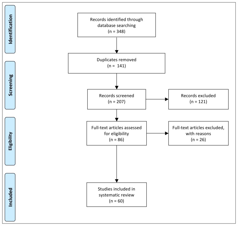
Methods: A systematic research of PubMed, Ovid, and Cochrane Library electronic databases was performed according to PRISMA guideline. Two Authors searched and evaluated the articles independently; a third Author was involved to solve any disagreement. The Oxford Level of Evidence (LoE) was used to assess each article.
Results: There is an increasing interest in the application of SE in the evaluation of healthy and diseased tendons. Many different tendons are amenable for SE evaluation, such as the Achilles and patellar tendons, rotator cuff, common extensor tendons, quadriceps tendon, and the plantar fascia.
Conclusion: SE appears to be a very useful diagnostic tool, in particular in tendon pathology. This is a dynamic examination, provides an immediate evaluation of the tissue elasticity, and may be useful in recognizing tendon abnormalities and in implementing the information available with conventional US.
Level of evidence: IV.
Keywords: elastography; elastosonography; epicondylitis; plantar fasciitis; tendinopathy; tendon healing.
Figures


References
-
- Woo SL. Mechanical properties of tendons and ligaments. I. Quasi-static and nonlinear viscoelastic properties. Biorheology. 1982;19(3):385–396. - PubMed
-
- Woo SL, Fisher MB, Feola AJ. Contribution of biomechanics to management of ligament and tendon injuries. Molecular & cellular biomechanics: MCB. 2008;5(1):49–68. - PubMed
-
- Woo SL, Orlando CA, Camp JF, Akeson WH. Effects of postmortem storage by freezing on ligament tensile behavior. Journal of biomechanics. 1986;19(5):399–404. - PubMed
-
- Giuseppetti GM, Martegani A, Di Cioccio B, Baldassarre S. Elastosonography in the diagnosis of the nodular breast lesions: preliminary report. La Radiologia medica. 2005;110(1–2):69–76. - PubMed
LinkOut - more resources
Full Text Sources
Other Literature Sources