. 2017:2017:8272504.
doi: 10.1155/2017/8272504. Epub 2017 Dec 13.

Neutrophil Extracellular Traps Are Pathogenic in Ventilator-Induced Lung Injury and Partially Dependent on TLR4

Affiliations

Neutrophil Extracellular Traps Are Pathogenic in Ventilator-Induced Lung Injury and Partially Dependent on TLR4

Haosi Li et al. Biomed Res Int. 2017.

Abstract

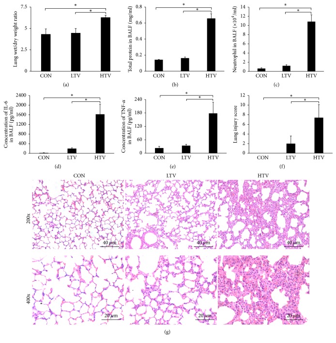
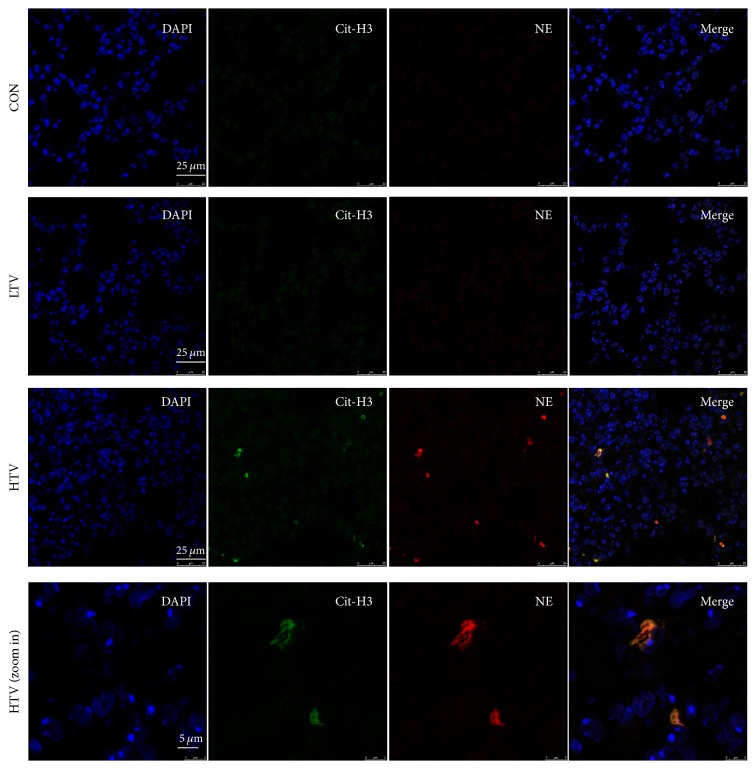
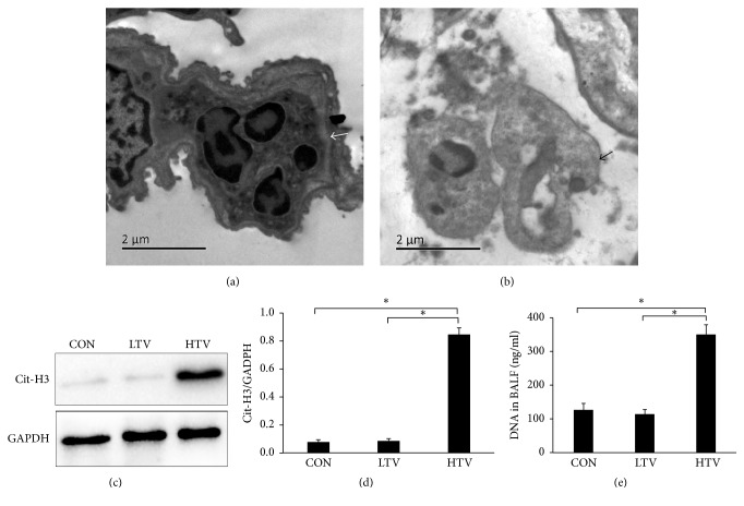
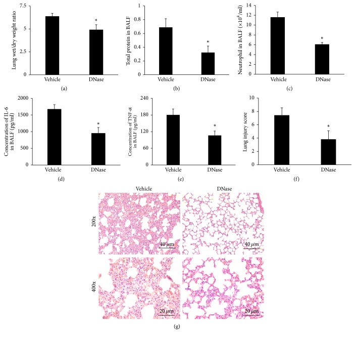
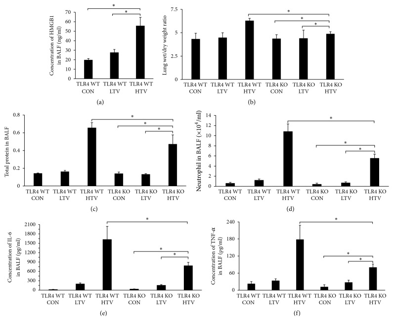
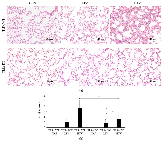
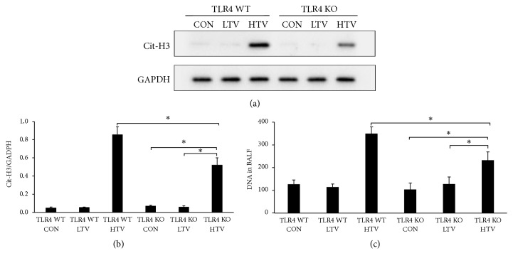
The pathogenesis of ventilator-induced lung injury (VILI) is associated with neutrophils. Neutrophils release neutrophil extracellular traps (NETs), which are composed of DNA and granular proteins. However, the role of NETs in VILI remains incompletely understood. Normal saline and deoxyribonuclease (DNase) were used to study the role of NETs in VILI. To further determine the role of Toll-like receptor 4 (TLR4) in NETosis, we evaluated the lung injury and NET formation in TLR4 knockout mice and wild-type mice that were mechanically ventilated. Some measures of lung injury and the NETs markers were significantly increased in the VILI group. DNase treatment markedly reduced NETs markers and lung injury. After high-tidal mechanical ventilation, the NETs markers in the TLR4 KO mice were significantly lower than in the WT mice. These data suggest that NETs are generated in VILI and pathogenic in a mouse model of VILI, and their formation is partially dependent on TLR4.


Figures

Figure 1
Figure 1
Ventilation with high-tidal volume causes lung injury. (a) The lung wet-to-dry weight ratio was increased in the HTV group compared with the CON group and the LTV group. (b) The concentration of the total protein in the BALF was significantly elevated in the HTV mice compared to the CON mice and the LTV mice. (c) The number of neutrophils in the BALF was increased in the HTV group. (d)(e) The BALF levels of IL-6 and TNF-α were markedly elevated in the HTV mice compared with the two other groups. (f) The lung injury score in the HTV group was markedly elevated compared to the other groups. (g) Low (magnification 200x, scale bar = 40 μm) and high power (magnification 400x, scale bar = 20 μm) views of the lungs of mice subjected to spontaneous ventilation, protective ventilation, and injurious ventilation. Hematoxylin and eosin stain. CON group: there are no obvious histological changes in the control group; the HTV group mice have much more inflammatory cell infiltration, alveolar septal thickening, pulmonary edema, and hemorrhage than in the CON and LTV groups. All the data are presented as the mean ± SD. P < 0.05.
Figure 2
Figure 2
Immunofluorescence staining of Cit-H3 and NE on lung sections. NETs, defined as colocalized DNA (blue), citrullinated histone-3 (Cit-H3) (green), and neutrophil elastase (NE) (red), presented in lungs sections of VILI model (HTV). However, there was no obvious staining in CON group and LTV group.
Figure 3
Figure 3
NETs formed in the lung of the VILI animal model. (a) Normal neutrophils in the airway with an intact nuclear membrane, located in the alveolar space, are shown by transmission electron micrograph (as indicated by the white arrows). Scale bar = 2 μm. (b) Neutrophils with a collapse of the nuclear membrane, dispersed chromatin, and nuclear vesicles (as indicated by the black arrows) in the alveolar space. Scale bar = 2 μm. (c) Representative in the lung homogenates. All the experiments were repeated at least 3 times. Only the HTV group has a strong Cit-H3 band. (d) The ratio of the Cit-H3/GAPDH intensity is significantly higher in the HTV group compared to the other groups. (e) The HTV mice produced a remarkably high level of BALF free DNA compared to the CON and LTV groups. P < 0.05.
Figure 4
Figure 4
DNase I treatment abolished NET formation in vivo. (a) The DNase I pretreated mice had a significantly decreased level of Cit-H3 expression in the lung homogenates by a Western analysis compared to the vehicle group. (b) The ratio of the Cit-H3/GAPDH intensity was significantly lower in the DNase group compared to the vehicle groups. (c) The DNase treated mice produced a lower level of BALF DNA compared to the vehicle group. P < 0.05 compared to the vehicle group.
Figure 5
Figure 5
DNase attenuates lung injury and inflammation in mice. (a) The lung wet/dry weight ratio decreased partially in the DNase group compared with the vehicle group. (b)(c) The BALF total protein concentration and the neutrophil counts in the BALF all decreased in the DNase I pretreated mice (versus vehicle control). (d)(e) DNase I also reduced the BALF levels of IL-6 and TNF-α. (f) The lung injury score in the vehicle group was markedly higher than those in the DNase group. (g) Low (magnification 200x, scale bar = 40 μm) and high power (magnification 400x, scale bar = 20 μm) views of the lungs of the vehicle group and the DNase group. Hematoxylin and eosin stain. The vehicle group had much more inflammatory cell infiltration, alveolar septal thickening, pulmonary edema, and hemorrhage than the DNase group. P < 0.05 compared to the vehicle group.
Figure 6
Figure 6
Toll-like receptor 4 knockout prevents the development of lung injury (A). (a) The level of HMGB1 in BALF was significantly higher than that in TLR4 WT CON group and LTV group. (b) The lung wet/dry ratio in TLR4 KO HTV mice was decreased compared with the TLR4 WT mice following HTV. (c)(d) The TLR4 KO HTV mice showed a marked elevation in protein content and neutrophil counts in the BALF compared to the TLR4 WT HTV mice. (e)(f) The BALF levels of IL-6 and TNF-α in the TLR4 KO HTV mice did not increase as much as in the TLR4 WT HTV mice. From (b) to (f), two-way ANOVA indicated both TLR4 genotype (TLR4 WT versus KO) and ventilation were significant, but there was no interaction (P < 0.05). P < 0.05.
Figure 7
Figure 7
Toll-like receptor 4 knockout prevents the development of lung injury (B). (a) Low (magnification 200x, scale bar = 40 μm) power views of the lungs of spontaneous ventilation, protective ventilation, and injurious ventilation in the TLR4 WT mice and the TLR4 KO mice, respectively. Hematoxylin and eosin stain. The TLR4 KO mice have less inflammatory cell infiltration, alveolar septal thickening, pulmonary edema, and hemorrhage than the TLR4 WT mice during HTV. (b) The lung injury score in the TLR4 KO HTV group was significantly lower than those in the TLR4 WT HTV group. Two-way ANOVA indicated that only ventilation was significant (P>0.05), but analyzing TLR4 WT HTV and TLR4 KO HTV with Student's t-test indicated a statistical significance (P > 0.05). P < 0.05.
Figure 8
Figure 8
Toll-like receptor 4 contributes to the NET formation in VILI in mice. (a) In contrast to the TLR4 WT mice, HTV partially increased Cit-H3 expression in the lung homogenates in the TLR4 KO mice. (b) The ratio of the Cit-H3/GAPDH intensity in the TLR4 KO HTV group was lower compared to the TLR4 WT HTV mice. Two-way ANOVA indicated that only ventilation was significant (P < 0.05), but analyzing TLR4 WT HTV and TLR4 KO HTV with Student's t-test indicated a statistical significance (P < 0.05). (c) The release of DNA in the BALF was markedly decreased in the TLR4 KO mice compared with the TLR4 WT mice after 4 h of ventilation. Two-way ANOVA indicated both TLR4 genotype and ventilation were significant, but there was no interaction (P < 0.05). P < 0.05.

Similar articles

Cited by

References

    1. Villar J., Blanco J., Añón J. M., et al. The ALIEN study: incidence and outcome of acute respiratory distress syndrome in the era of lung protective ventilation. Intensive Care Medicine. 2011;37(12):1932–1941. doi: 10.1007/s00134-011-2380-4. - DOI - PubMed
    1. Imai Y., Kawano T., Miyasaka K., Takata M., Imai T., Okuyama K. Inflammatory chemical mediators during conventional ventilation and during high frequency oscillatory ventilation. American Journal of Respiratory and Critical Care Medicine. 1994;150(6 I):1550–1554. doi: 10.1164/ajrccm.150.6.7952613. - DOI - PubMed
    1. Tremblay L., Valenza F., Ribeiro S. P., Li J., Slutsky A. S. Injurious ventilatory strategies increase cytokines and c-fos m-RNA expression in an isolated rat lung model. The Journal of Clinical Investigation. 1997;99(5):944–952. doi: 10.1172/JCI119259. - DOI - PMC - PubMed
    1. Takata M., Abe J., Tanaka H., et al. Intraalveolar expression of tumor necrosis factor-α gene during conventional and high-frequency ventilation. American Journal of Respiratory and Critical Care Medicine. 1997;156(1):272–279. doi: 10.1164/ajrccm.156.1.9607072. - DOI - PubMed
    1. Imanaka H., Shimaoka M., Matsuura N., Nishimura M., Ohta N., Kiyono H. Ventilator-induced lung injury is associated with neutrophil infiltration, macrophage activation, and TGF-β1 mRNA upregulation in rat lungs. Anesthesia & Analgesia. 2001;92(2):428–436. doi: 10.1213/00000539-200102000-00029. - DOI - PubMed

MeSH terms

LinkOut - more resources