. 2018 Jan 28:13:83-92.
doi: 10.1016/j.bbrep.2018.01.005. eCollection 2018 Mar.

Involvement of the ubiquitin-proteasome system in the expression of extracellular matrix genes in retinal pigment epithelial cells

Affiliations

Involvement of the ubiquitin-proteasome system in the expression of extracellular matrix genes in retinal pigment epithelial cells

J Emanuel Ramos de Carvalho et al. Biochem Biophys Rep. .

Abstract

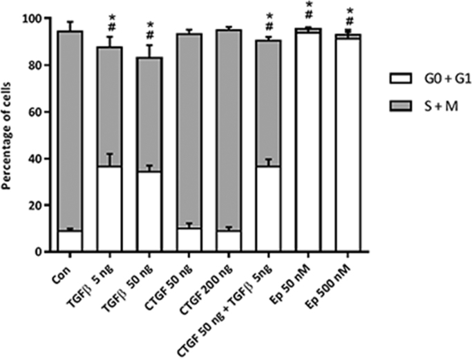
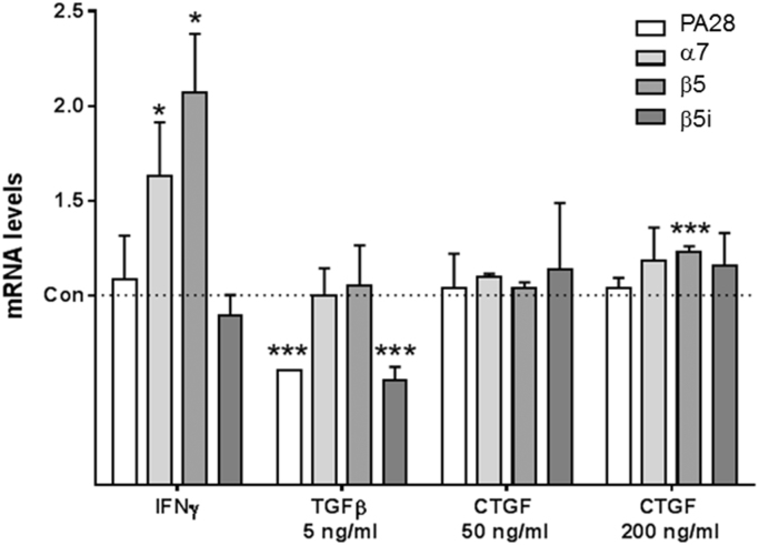
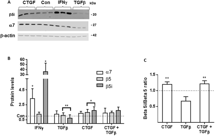
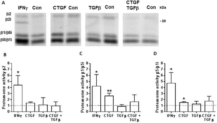
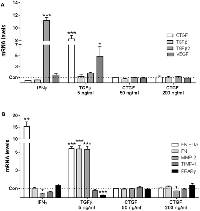
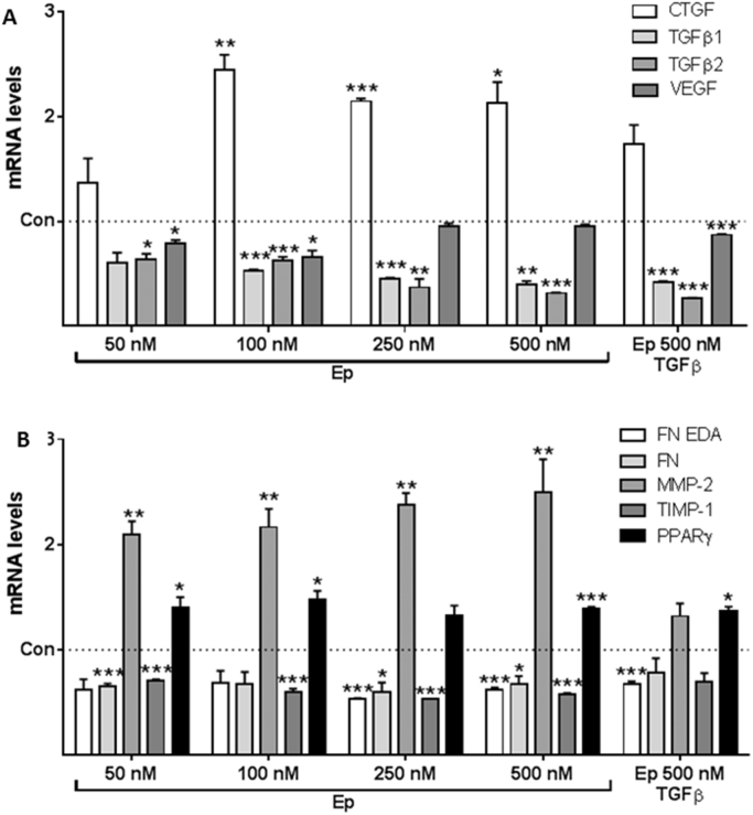
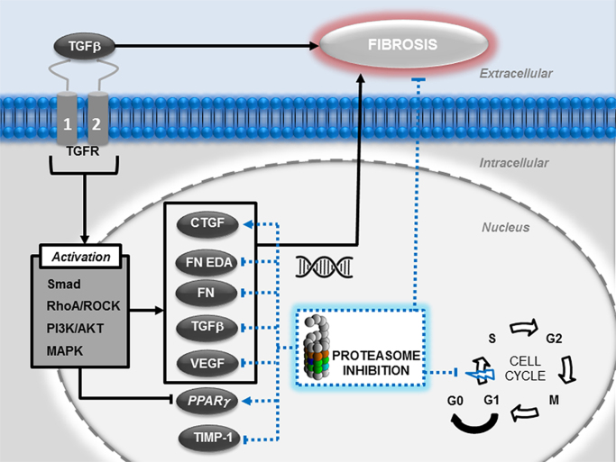
Emerging evidence suggests that dysfunction of the ubiquitin-proteasome system is involved in the pathogenesis of numerous senile degenerative diseases including retinal disorders. The aim of this study was to assess whether there is a link between proteasome regulation and retinal pigment epithelium (RPE)-mediated expression of extracellular matrix genes. For this purpose, human retinal pigment epithelial cells (ARPE-19) were treated with different concentrations of transforming growth factor-β (TGFβ), connective tissue growth factor (CTGF), interferon-γ (IFNγ) and the irreversible proteasome inhibitor epoxomicin. First, cytotoxicity and proliferation assays were carried out. The expression of proteasome-related genes and proteins was assessed and proteasome activity was determined. Then, expression of fibrosis-associated factors fibronectin (FN), fibronectin EDA domain (FN EDA), metalloproteinase-2 (MMP-2), tissue inhibitor of metalloproteinases-1 (TIMP-1) and peroxisome proliferator-associated receptor-γ (PPARγ) was assessed. The proteasome inhibitor epoxomicin strongly arrested cell cycle progression and down-regulated TGFβ gene expression, which in turn was shown to induce expression of pro-fibrogenic genes in ARPE-19 cells. Furthermore, epoxomicin induced a directional shift in the balance between MMP-2 and TIMP-1 and was associated with down-regulation of transcription of extracellular matrix genes FN and FN-EDA and up-regulation of the anti-fibrogenic factor PPARγ. In addition, both CTGF and TGFβ were shown to affect expression of proteasome-associated mRNA and protein levels. Our results suggest a link between proteasome activity and pro-fibrogenic mechanisms in the RPE, which could imply a role for proteasome-modulating agents in the treatment of retinal disorders characterized by RPE-mediated fibrogenic responses.

Keywords: AMD, age-related macular degeneration; ARPE-19, human retinal pigment epithelial cells; CNV, choroidal neovascularization; CTGF; CTGF, connective tissue growth factor; ECM, extracellular matrix; EMT, epithelial-mesenchymal transition; Epoxomicin; FN EDA, fibronectin EDA domain; FN, fibronectin; Fibrosis; IFNγ, interferon-γ; MMP-2, matrix metalloproteinase-2; PPARγ; PPARγ, peroxisome proliferator-associated receptor-γ; Proteasome; RPE; RPE, retinal pigment epithelium; Retina; TGFβ; TGFβ, transforming growth factor-β; TIMP-1, tissue inhibitor of metalloproteinases-1; UPS, ubiquitin-proteasome system; nAMD, neovascular age-related macular degeneration.


Figures

Fig. 1
Fig. 1
Proliferation of ARPE-19 cells in the presence of TGFβ, CTGF, TGFβ and CTGF or epoxomicin expressed as percentage of cells in the S and M phase versus cells in the G0 and G1 phase after flow cytometric analysis of the percentage of cells that had incorporated EdU. *Significant difference from control of percentage of cells in G0 and G1 phase. #Significant difference from control of percentage of cells in the S and M phase. The experiment was performed in triplicate and repeated twice (N = 2).
Fig. 2
Fig. 2
Proteasome mRNA levels induced by IFNγ, TGFβ or CTGF. mRNA levels of PA28α, β5, β5i, and α7 subunits of the proteasome in ARPE-19 cells, after stimulation with IFNγ, TGFβ and low and high concentrations of CTGF. IFNγ upregulates mRNA expression of α7 and β5 subunits whereas TGFβ downregulates mRNA expression of PA28α and β5i subunits and CTGF upregulates mRNA expression of β5 subunit. Values represent mRNA expression levels (mean ± SD) relative to untreated control cells. *, Significant change (P < 0.05); ***, significant change (P < 0.001). The experiment was performed in triplicate (N = 3).
Fig. 3
Fig. 3
Proteasome-specific subunit protein expression and β5i:β5 ratios upon IFNγ, CTGF, TGFβ or CTGF and TGFβ stimulation of ARPE-19 cells. (A) Western blot showing protein levels of β5i, α7 and actin (loading control) in cells that had been incubated with IFNγ, CTGF or TGFβ. (B) Protein levels of β5 and β5i subunits were assessed by western blot with actin as loading control and α7 subunit as proteasome content control. (C) Quantitative data of the average ratio of β5i and β5 relative to control samples after incubation in the presence or absence of CTGF, TGFβ or CTGF and TGFβ. Data are expressed as the mean ± SD. *, Significant change (P < 0.05): **, significant change (P < 0.01). The experiment was performed in triplicate (N = 3).
Fig. 4
Fig. 4
Increased specific proteasome activity upon CTGF stimulation of ARPE-19 cells. (A) After treatment with IFNγ (50 U), TGFβ (50 ng), CTGF (50 ng) or CTGF (50 ng) followed 6 h later by TGFβ (5 ng), ARPE-19 cells were harvested and proteasomes were labeled with a Bodipy-Ep activity probe. Proteasome activities were assessed by western blotting. Quantitative data of the proteasome activity are presented for proteasome subunit β2 (B), β1/β5i complex (C), and β5/β1i complex (D). *, Significant change (P < 0.05); **, significant change (P < 0.01). The experiment was performed in triplicate (N = 3).
Fig. 5
Fig. 5
TGFβ upregulates mRNA expression of CTGF, VEGF and pro-fibrogenic genes and downregulates mRNA expression of the anti-fibrogenic factor PPARγ. After stimulation with TGFβ, CTGF and IFNγ, mRNA levels of (A) CTGF, TGFβ1, TGFβ2, VEGF and (B) FN, FN EDA, MMP-2, TIMP-1 and PPARγ were assessed in ARPE-19 cells. Values represent mRNA expression levels (mean ± SD) relative to untreated control cells. *, Significant change (P < 0.05); **, significant change (P < 0.01); ***, significant change (P < 0.001). The experiment was performed in triplicate (N = 3).
Fig. 6
Fig. 6
Epoxomicin (Ep) downregulates mRNA expression of TGFβ1, TGFβ2, VEGF, FN, FN EDA, TIMP-1 and upregulates mRNA expression of CTGF and PPARγ. In the presence of TGFβ, epoxomicin dowregulates mRNA expression of TGFβ1, TGFβ2, VEGF and FN EDA and expression of PPARγ is up-regulated. After treatment with increasing concentrations of epoxomicin in untreated and TGFβ-treated ARPE-19 cells, mRNA levels of (A) CTGF, TGFβ1, TGFβ2, VEGF, (B) FN, FN EDA, MMP-2, TIMP-1 and PPARγ were assessed. Values represent mRNA expression levels (mean ± SD) relative to untreated control cells. *, Significant change (P < 0.05); **, significant change (P < 0.01); ***, significant change (P < 0.001). The experiment was performed in triplicate (N = 3).
Fig. 7
Fig. 7
A role for proteasome inhibition in the modulation of fibrogenic mechanisms mediated by RPE cells. TGFβ activates multiple pathways, including the Smad, Rho-like GTPase, PI3K/AKT and MAPK pathways, resulting in the transcription of several pro-fibrogenic genes such as CTGF, FN, FN EDA, VEGF and down-regulation of PPARγ transcription. These effects contribute to epithelial-mesenchymal transition processes in RPE cells and initiation of fibrosis. Proteasome inhibition halts cell cycle progression and downregulates transcription of FN, FN EDA, TGFβ, VEGF and TIMP-1 whilst transcription of the anti-fibrogenic factor PPARγ is up-regulated, also upon exposure to TGFβ. Effects of proteasome inhibition are depicted as dotted lines. Abbreviations: connective tissue growth factor (CTGF); fibronectin (FN); fibronectin EDA (FN EDA); MAP kinase pathway (MAPK); phosphatidylinositol-3-kinase pathway (PI3K/AKT); retinal pigment epithelium cells (RPE); Rho-like GTPase pathway (RhoA/ROCK); tissue inhibitor metalloproteinase-1 (TIMP-1); transforming growth factor β (TGFβ); transforming growth factor receptor (TGFR); vascular endothelial growth factor (VEGF).

Similar articles

Cited by

References

    1. Augood C.A., Vingerling J.R., De Jong P.T.V.M., Chakravarthy U., Seland J., Soubrane G., Tomazzoli L., Topouzis F., Bentham G., Rahu M., Vioque J., Young I.S., Fletcher A.E. Prevalence of age-related maculopathy in older Europeans - the European Eye Study (EUREYE) Arch. Ophthalmol. 2006;124:529–535. - PubMed
    1. Spaide R.F., Curcio C.A. Drusen characterization with multimodal imaging. Retina. 2010;30:1441–1454. - PMC - PubMed
    1. Crabb J.W., Miyagi M., Gu X.R., Shadrach K., West K.A., Sakaguchi H., Kamei M., Hasan A., Yan L., Rayborn M.E., Salomon R.G., Hollyfield J.G. Drusen proteome analysis: an approach to the etiology of age-related macular degeneration. Proc. Natl. Acad. Sci. USA. 2002;99:14682–14687. - PMC - PubMed
    1. Nagai N., Klimava A., Lee W.H., Izumi-Nagai K., Handa J.T. CTGF is increased in basal deposits and regulates matrix production through the ERK (p42/p44(mapk)) MAPK and the p38 MAPK signaling pathways. Investig. Ophthalmol. Vis. Sci. 2009;50:1903–1910. - PMC - PubMed
    1. Klein R., Chou C.F., Klein B.E.K., Zhang X.Z., Meuer S.M., Saaddine J.B. Prevalence of age-related macular degeneration in the US population. Arch. Ophthalmol. 2011;129:75–80. - PubMed