Review
. 2018 Apr;9(2):121-135.
doi: 10.1007/s13244-018-0593-6. Epub 2018 Jan 31.

Imaging of non-neoplastic duodenal diseases. A pictorial review with emphasis on MDCT

Affiliations
Review

Imaging of non-neoplastic duodenal diseases. A pictorial review with emphasis on MDCT

Sergi Juanpere et al. Insights Imaging. 2018 Apr.

Abstract

A wide spectrum of abnormalities can affect the duodenum, ranging from congenital anomalies to traumatic and inflammatory entities. The location of the duodenum and its close relationship with other organs make it easy to miss or misinterpret duodenal abnormalities on cross-sectional imaging. Endoscopy has largely supplanted fluoroscopy for the assessment of the duodenal lumen. Cross-sectional imaging modalities, especially multidetector computed tomography (MDCT) and magnetic resonance imaging (MRI), enable comprehensive assessment of the duodenum and surrounding viscera. Although overlapping imaging findings can make it difficult to differentiate between some lesions, characteristic features may suggest a specific diagnosis in some cases. Familiarity with pathologic conditions that can affect the duodenum and with the optimal MDCT and MRI techniques for studying them can help ensure diagnostic accuracy in duodenal diseases. The goal of this pictorial review is to illustrate the most common non-malignant duodenal processes. Special emphasis is placed on MDCT features and their endoscopic correlation as well as on avoiding the most common pitfalls in the evaluation of the duodenum.

Teaching points: • Cross-sectional imaging modalities enable comprehensive assessment of duodenum diseases. • Causes of duodenal obstruction include intraluminal masses, inflammation and hematomas. • Distinguishing between tumour and groove pancreatitis can be challenging by cross-sectional imaging. • Infectious diseases of the duodenum are difficult to diagnose, as the findings are not specific. • The most common cause of nonvariceal upper gastrointestinal bleeding is peptic ulcer disease.

Keywords: Duodenal lesions; Duodenum; Imaging; MDCT; MRI.


Figures

Fig. 1
Fig. 1
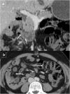
Duodenal duplication cyst. Reformatted coronal contrast-enhanced CT image shows a large well-circumscribed cystic mass (arrows) adjacent to the second duodenal portion. There is no communication with the duodenal lumen (*)
Fig. 2
Fig. 2
Ruptured duodenal duplication cyst in a 58-year-old man who sustained blunt trauma in a fall down stairs. a Reformatted sagittal contrast-enhanced CT shows circumferential wall thickening of the cyst and a disruption in its anterior surface (arrow). b Axial CT shows retroperitoneal fluid in the right anterior pararenal space (white arrow). The duodenal lumen is narrowed and displaced medially due to inflammatory changes (black arrow). c Axial T2-weighted MRI shows stranding of the periduodenal fat on (arrows). d Axial T1-weighted MRI image shows a hyperintensity of the content of the lesion (*). The high signal intensity (more than muscle tissue), in a traumatic context, suggests an haemorrhagic transformation of the cyst. e Detail of the fiberoptic endoscopic study
Fig. 3
Fig. 3
Duodenal diverticula in different patients. a Anteroposterior image from a single-contrast upper gastrointestinal series shows a typical outpouching (*) arising from descending duodenum. b Magnetic resonance cholangiopancreatography shows a heterogeneous outpouching (large arrow) in the papillary region. Note common bile duct (arrow) and proximal Wirsung duct (arrowhead). c Axial CT demonstrates two duodenal diverticula (arrows) in the periampullary region (air-filled) and fourth portion of the duodenum (with air-fluid level). d Axial CT reveals a diverticulum (arrow) that contains debris in the third portion of the duodenum. Air bubbles inside the lesion are virtually pathognomonic for a duodenal diverticulum
Fig. 4
Fig. 4
A 67-year-old man with epigastralgia, fever, and food intolerance. a Initial abdominal ultrasonography shows diffuse thickening of the different layers of the duodenal wall with a slight amount of free periduodenal fluid (arrows) and an adjacent hypoechoic fluid collection (*). b Axial contrast-enhanced CT confirms the presence of a paraduodenal collection with an air-fluid level (*) and surrounding soft-tissue stranding (arrows) caused by acute duodenal diverticulitis. c Axial T2-weighted MRI after conservative therapy shows the outpouching from the duodenal wall is smaller and more well-defined; it now has the appearance of a duodenal diverticulum, and the amount of soft-tissue stranding has decreased
Fig. 5
Fig. 5
Intraduodenal diverticulum in a 58-year-old woman. Single-contrast upper gastrointestinal study in the right anterior oblique projection shows a well-defined oval lesion (arrow) surrounded by a radiotransparent halo (“windsock” sign). The lesion contains oral contrast material and projects into the true duodenal lumen
Fig. 6
Fig. 6
Gastrointestinal nonrotation in a 46-year-old woman. a Reformatted coronal contrast-enhanced CT image shows that the third portion of the duodenum (arrows) does not cross the midline of the abdomen. b The small bowel is located in the right side of the abdomen and the colon in the left side (arrows) on axial CT image
Fig. 7
Fig. 7
Annular pancreas in a 43-year-old man with a long history of abdominal pain. Contrast-enhanced CT shows the pancreas (arrows) completely encircling the descending duodenum (*)
Fig. 8
Fig. 8
Spectrum of duodenitis in different patients. a Axial contrast-enhanced CT shows thickened oedematous duodenal wall with inflammation involving periduodenal fat (arrows) in nonspecific duodenitis. b Reformatted coronal contrast-enhanced CT image in a woman with disseminated breast cancer and secondary hepatic insufficiency shows nonspecific mural thickening of the duodenum with mucosal enhancement (arrows)
Fig. 9
Fig. 9
Tuberculosis in a 63-year-old man with constitutional symptoms, fever, and abdominal pain. Axial contrast-enhanced CT shows a masslike appearance (white arrow) that encompasses the superior mesenteric artery and a fistulous pathway that communicates with the duodenal lumen (black arrow). Note the marked extrinsic compression of the third portion of the duodenum. CT also shows periduodenal fat stranding (short arrows) and a necrotic lymph node (arrowhead)
Fig. 10
Fig. 10
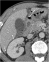
A 48-year-old man who presented with abdominal pain caused by a perforated duodenal ulcer. Axial contrast-enhanced CT shows an irregular air-fluid level (large arrow) adjacent to the duodenal bulb. A leakage of free fluid into the posterior subhepatic space (white arrows) through hepatoduodenal ligament (black arrows) is observed
Fig. 11
Fig. 11
An 88-year-old man who was treated with oral anticoagulants for ischemic heart disease, presented with coffee ground vomitus. Axial contrast-enhanced CT shows homogeneous concentric thickening (arrows) of the duodenal wall at the level of the bulb. Upper endoscopy detected an ulcer in the duodenal bulb with active oozing haemorrhage which was sclerosed with adrenaline
Fig. 12
Fig. 12
A 63-year-old male smoker with upper digestive bleeding. Initial upper endoscopy showed a large bleeding ulcer on the anterior surface of the duodenal bulb; sclerosing therapy with adrenaline and aethoxysklerol® was unsuccessful. a Arteriogram shows contrast medium extravasated from the posterior pancreaticoduodenal arcade (arrows) into the duodenum. b Coil embolization of gastroduodenal artery stopped the bleeding
Fig. 13
Fig. 13
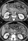
Acute exsudative pancreatitis and duodenal edema in a 50-year-old man. Contrast-enhanced CT shows an enlarged pancreatic head (*) with stranding of the peripancreatic fat and peripancreatic fluid (short arrows). The wall of the duodenum is thickened, and limited mural enhancement is seen secondary to edema (arrows). Also note the pathologic amount of fluid in the upper abdomen (curved arrows)
Fig. 14
Fig. 14
Duodenal compression by pancreatic pseudocyst in a 69-year-old man. a Axial contrast-enhanced CT reveals lateral displacement of the descending duodenum (arrows) by a pancreatic fluid collection (*). b Detail of an internal drain (arrow) guided by echoendoscopy to improve secondary food intolerance
Fig. 15
Fig. 15
A 48-year-old man with epigastric pain. Contrast-enhanced CT identifies subtle infiltration of soft tissue (white arrow) in the pancreaticoduodenal groove. Duodenal mural thickening secondary to edema is observed on the medial side (black arrow). Note the normal enhancement of the pancreatic head and absence of classic signs of exsudative pancreatitis
Fig. 16
Fig. 16
A 55-year-old man with history of alcohol abuse and chronic pancreatitis. Axial contrast-enhanced CT images (a and b) demonstrate atrophy of the pancreatic parenchyma and ductal dilation (short arrow). A stricture of the duodenal lumen (arrow) caused by fibrotic changes in the pancreaticoduodenal groove is accompanied by gastric dilation (*). c Image from a barium study shows a stricture in the proximal duodenum with an abnormal mucosal pattern (arrow)
Fig. 17
Fig. 17
A 56-year-old man with history of alcohol abuse and smoking presented with weight loss. a Axial contrast-enhanced CT reveals cystic dystrophy with multiple cystic parietal areas (arrows) in the thickened second duodenal portion causing severe duodenal obstruction with gastric dilation (*). Atrophy of the pancreatic parenchyma and strictures of the pancreatic duct (short arrow) due to chronic pancreatitis. b Detail from the upper gastrointestinal endoscopy study showing extrinsic compression of the duodenal lumen
Fig. 18
Fig. 18
A 24-year-old man with Crohn’s disease who presented with reflux esophagitis, daily vomiting, and food intolerance. a Upper endoscopy shows pyloric and bulbar involvement with ulcerated areas and folded hypertrophy that caused stenosis. After the dilation of the stenosis b the patient presented acute abdominal pain. Axial c and reformatted sagittal d contrast-enhanced CT images confirm the suspected perforation (short arrows) and also show the thickened and hyperaemic wall of the duodenum (arrows) with fibrotic bulbar stenosis
Fig. 19
Fig. 19
Acute cholecystitis in a 54-year-old man. a Ultrasonography identified an enlargement of the lateral wall of the duodenum (white arrows) that was accompanied by a periduodenal fluid collection (*). b Reformatted coronal contrast-enhanced CT image shows a distended gallbladder with inaccurate margins (black arrow) and confirms the presence of a periduodenal fluid collection (*). Also note the secondary duodenal wall involvement (white arrow)
Fig. 20
Fig. 20
Right upper quadrant pain and gallstone ileus in a 39-year-old woman. Axial contrast-enhanced CT (a and b) shows air in the bile duct (arrow in a), a thick-walled gallbladder, and a fistula to the duodenum (arrow in b). Reformatted coronal contrast-enhanced CT image demonstrates two gallstones (arrows) within the proximal ileum with proximal jejunal dilation
Fig. 21
Fig. 21
Duodenal trauma in different patients. a A 20-year-old man with blunt abdominal trauma. Contrast-enhanced CT shows an extensive pancreaticoduodenal hematoma with displacement of the mesenteric vessels to the left (white arrow) and disruption of the wall of the third duodenal portion (black arrow) with active extravasation of intravenous contrast (arrowhead). Note the abundant retroperitoneal fluid (short arrow). b A 45-year-old man involved in a motor vehicle accident. Contrast-enhanced CT shows free retroperitoneal fluid (arrow) adjacent to the third duodenal portion and slight wall thickening or mural edema. Abdominal subcutaneous emphysema (short arrows) secondary to multiple rib fractures and pneumothorax
Fig. 22
Fig. 22
Intramural duodenal hematoma in a 28-year-old man with a history of cocaine abuse. a Unenhanced CT shows a spontaneous hyperdense thickening of the duodenal wall (arrow). b Axial contrast-enhanced CT shows a heterogeneous and intramural duodenal masslike appearance (arrow). c Detail of the endoscopic study in the initial diagnostic. d Reformatted coronal contrast-enhanced CT image of the same patient a few days before show a predominant hypodense expansive masslike appearance (arrow) causing a mass effect of the duodenal lumen with gastric dilation
Fig. 23
Fig. 23
A 58-year-old man with coffee ground vomitus, hypotension, and abdominal pain. Reformatted coronal unenhanced CT image shows a gastroparesis caused by extensive intramural bowel gas (pneumatosis intestinalis) that affects the entire stomach (black arrows) and duodenum (white arrows). Also note gas in the portal vein (double arrow). Upper endoscopy shows a necrotic gastric and duodenal mucosa
Fig. 24
Fig. 24
A 31-year-old woman with suspected superior mesenteric artery (SMA) syndrome. Axial contrast-enhanced CT images (a, b, and c) show a dilated proximal duodenum (*) with abrupt narrowing of the third portion (black arrows) corresponding with the course of SMA (white arrow). Reformatted sagittal contrast-enhanced CT image demonstrates compression of the third portion of the duodenum (black arrow) between the SMA (white arrow) and aorta

Similar articles

Cited by

References

    1. Jayaraman MV, Mayo-Smith WW, Movson JS et al (2001) CT of the duodenum; an overlooked segment gets its due. Radiographics 21 Spec No:S147–S160 - PubMed
    1. Barat M, Dohan A, et al. Mass-forming lesions of the duodenum a pictorial review. Diagn Interv Imaging. 2017;98(10):663–675. doi: 10.1016/j.diii.2017.01.004. - DOI - PubMed
    1. McNeeley MF, Lalwani N, et al. Multimodality imaging of diseases of the duodenum. Abdom Imaging. 2014;39:1330–1349. doi: 10.1007/s00261-014-0157-2. - DOI - PubMed
    1. Sun CH, Li X, et al. Multidetector computed tomography (MDCT) manifestations of the normal duodenal papilla. Eur J Radiol. 2013;82(6):918–922. doi: 10.1016/j.ejrad.2013.01.007. - DOI - PubMed
    1. Pearl MS, Hill MC, et al. CT findings in duodenal diverticulitis. AJR. 2006;187:W392–W395. doi: 10.2214/AJR.06.0215. - DOI - PubMed

LinkOut - more resources