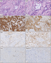
Clinicopathological characteristics and molecular analysis of primary pulmonary mucoepidermoid carcinoma: Case report and literature review
- PMID: 29388384
- PMCID: PMC5792747
- DOI: 10.1111/1759-7714.12565
Clinicopathological characteristics and molecular analysis of primary pulmonary mucoepidermoid carcinoma: Case report and literature review
Abstract
Primary pulmonary mucoepidermoid carcinoma (PMEC) is extremely rare. Herein, we report a case of a 71-year-old male patient with high-grade PMEC involving the right upper lobe that was successfully resected via lobectomy. As a result of invasion into the pleural and paratracheal lymph nodes, four cycles of adjuvant chemotherapy with paclitaxel and carboplatin were administered. There were no signs of relapse during 10 months of follow-up. Furthermore, we reviewed the literature and summarized the surgical approaches, prognostic factors, and underlying genetic mechanisms of PMEC, which will benefit clinical treatment.
Keywords: Chemotherapy; MECT1-MAML2 fusion gene; lobectomy; pulmonary mucoepidermoid carcinoma.
© 2017 The Authors. Thoracic Cancer published by China Lung Oncology Group and John Wiley & Sons Australia, Ltd.
Figures


Similar articles
-
A case of pulmonary mucoepidermoid carcinoma responding to carboplatin and paclitaxel.Jpn J Clin Oncol. 2014 May;44(5):493-6. doi: 10.1093/jjco/hyu016. Epub 2014 Mar 11. Jpn J Clin Oncol. 2014. PMID: 24620028
-
Primary Pulmonary Mucoepidermoid Carcinoma: Histopathological and Moleculargenetic Studies of 26 Cases.PLoS One. 2015 Nov 17;10(11):e0143169. doi: 10.1371/journal.pone.0143169. eCollection 2015. PLoS One. 2015. PMID: 26575266 Free PMC article.
-
Clinicopathologic and genetic features of primary bronchopulmonary mucoepidermoid carcinoma: the MD Anderson Cancer Center experience and comprehensive review of the literature.Virchows Arch. 2017 Jun;470(6):619-626. doi: 10.1007/s00428-017-2104-4. Epub 2017 Mar 25. Virchows Arch. 2017. PMID: 28343305 Review.
-
Pathological complete response to gefitinib in a 10-year-old boy with EGFR-negative pulmonary mucoepidermoid carcinoma: a case report and literature review.Clin Respir J. 2017 May;11(3):346-351. doi: 10.1111/crj.12343. Epub 2015 Aug 6. Clin Respir J. 2017. PMID: 26148572 Review.
-
Prognostic factors of primary pulmonary mucoepidermoid carcinoma: a clinical and pathological analysis of 34 cases.Int J Clin Exp Pathol. 2014 Sep 15;7(10):6792-9. eCollection 2014. Int J Clin Exp Pathol. 2014. PMID: 25400760 Free PMC article.
Cited by
-
Pulmonary Mucoepidermoid Carcinoma With Multiple Paraneoplastic Syndromes.Cureus. 2023 Aug 27;15(8):e44193. doi: 10.7759/cureus.44193. eCollection 2023 Aug. Cureus. 2023. PMID: 37767242 Free PMC article.
-
Left lower lobe sleeve resection for the clear cell variant of pulmonary mucoepidermoid carcinoma: A case report.World J Clin Cases. 2024 Mar 16;12(8):1422-1429. doi: 10.12998/wjcc.v12.i8.1422. World J Clin Cases. 2024. PMID: 38576804 Free PMC article.
-
Predictive CT features for the diagnosis of primary pulmonary mucoepidermoid carcinoma: comparison with squamous cell carcinomas and adenocarcinomas.Cancer Imaging. 2021 Jan 6;21(1):2. doi: 10.1186/s40644-020-00375-2. Cancer Imaging. 2021. PMID: 33407915 Free PMC article.
-
A surgical strategy-based nomogram for primary pulmonary mucoepidermoid carcinoma.Discov Oncol. 2025 Aug 15;16(1):1558. doi: 10.1007/s12672-025-03117-7. Discov Oncol. 2025. PMID: 40815387 Free PMC article.
-
Mucoepidermoid lung carcinoma in a pediatric patient confused with pneumonia.Radiol Case Rep. 2021 Jul 22;16(9):2749-2753. doi: 10.1016/j.radcr.2021.06.078. eCollection 2021 Sep. Radiol Case Rep. 2021. PMID: 34377224 Free PMC article.
References
Publication types
MeSH terms
Substances
LinkOut - more resources
Full Text Sources
Other Literature Sources
Medical
Research Materials

