Review
. 2018 May;55(3):140-162.
doi: 10.1080/10408363.2018.1425976. Epub 2018 Feb 1.

Advances in liquid biopsy on-chip for cancer management: Technologies, biomarkers, and clinical analysis

Affiliations
Review

Advances in liquid biopsy on-chip for cancer management: Technologies, biomarkers, and clinical analysis

Amogha Tadimety et al. Crit Rev Clin Lab Sci. 2018 May.

Abstract

Liquid biopsy, as a minimally invasive method of gleaning insight into the dynamics of diseases through a patient fluid sample, has been growing in popularity for cancer diagnosis, prognosis, and monitoring. While many technologies have been developed and validated in research laboratories, there has also been a push to expand these technologies into other clinical settings and as point of care devices. In this article, we discuss and evaluate microchip-based technologies for circulating tumor cell (CTC), exosome, and circulating tumor nucleic acid (ctNA) capture, detection, and analysis. Such integrated systems streamline otherwise multiple-step, manual operations to get a sample-to-answer quantitation. In addition, analysis of disease biomarkers is suited to point of care settings because of ease of use, low consumption of sample and reagents, and high throughput. We also cover the basics of biomarkers and their detection in biological fluid samples suitable for liquid biopsy on-chip. We focus on emerging technologies that process a small patient sample with high spatial-temporal resolution and derive clinically meaningful results through on-chip biomarker sensing and downstream molecular analysis in a simple workflow. This critical review is meant as a resource for those interested in developing technologies for capture, detection, and analysis platforms for liquid biopsy in a variety of settings.

Keywords: Liquid biopsy; cancer biomarker; biosensor; lab-on-chip; point of care.


Figures

Figure 1:
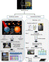
Figure 1:. Overview of Liquid Biopsies on-chip.
An overview of integrated capture and analysis of biomarkers for point of care cancer diagnosis. Adapted in part from A. Tadimety, A. Syed, Y. Nie, C. R. Long, K. M. Kready, and X. J. Zhang, “Liquid biopsy on chip: a paradigm shift towards the understanding of cancer metastasis,” Integr. Biol., vol. 64, pp. 9–29, 2017 with permission of The Royal Society of Chemistry[1].
Figure 2.
Figure 2.
Advantages of biosensor and Lab on Chip for liquid biopsy, including integration, miniaturization, and clinical advantages.
Figure 3.
Figure 3.
Typical circulating biomarkers and applications of their capture and analysis to diagnostics, prognostics, and therapy. Adapted with permission under the Creative Commons Attribution License 3.0 from S. Jia, R. Zhang, Z. Li, and J. Li, “Clinical and biological significance of CTCs, circulating tumor DNA (ctDNA), and exosomes as biomarkers in colorectal cancer,” Oncotarget, vol. 8, no. 33, pp. 55632–55645, 2015 [22].
Figure 4.
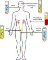
Figure 4.
Sample fluid types for liquid biopsy, including blood and its derivatives, cerebrospinal fluid, saliva, and urine. This figure is adapted with permission under the Creative Commons License 4.0 from A. Di Meo, J. Bartlett, Y. Cheng, M. D. Pasic, and G. M. Yousef, “Liquid biopsy: a step forward towards precision medicine in urologic malignancies,” Mol. Cancer, vol. 16, no. 1, p. 80, 2017 [47].
Figure 5.
Figure 5.
Magnetic sifter chip for capture and downstream analysis of CTCs using immunomagnetic separation and mutational analysis. Reproduced from C. M. Earhart, C. E. Hughes, R. S. Gaster, C. C. Ooi, R. J. Wilson, L. Y. Zhou, E. W. Humke, L. Xu, D. J. Wong, S. B. Willingham, E. J. Schwartz, I. L. Weissman, S. S. Jeffrey, J. W. Neal, R. Rohatgi, H. A. Wakelee, and S. X. Wang, “Isolation and mutational analysis of CTCs from lung cancer patients with magnetic sifters and biochips,” Lab Chip, vol. 14, no. 1, pp. 78–88, 2014 with permission of the Royal Society of Chemistry [61].
Figure 6.
Figure 6.. Immunomagnetic microchip and platform for CTC separation.
Top row: Picture of the device, schematic of operation, and images of captured cells. Reproduced from K. Hoshino, Y.-Y. Huang, N. Lane, M. Huebschman, J. W. Uhr, E. P. Frenkel, and X.J. Zhang, “Microchip-based immunomagnetic detection of CTCs,” Lab Chip, vol. 11, no. 20, p. 3449, 2011 with permission of the Royal Society of Chemistry [84]. Bottom row: commercial NanoLite CellRich™ Systems with embedded microfluidic cartridge developed based on the prototype devices.
Figure 7:
Figure 7:
Integrated nanoplatform for single CTC seeding and mutational analysis. Reproduced with permission from S. Park, D. J. Wong, C. C. Ooi, D. M. Kurtz, O. Vermesh, A. Aalipour, S. Suh, K. L. Pian, J. J. Chabon, S. H. Lee, M. Jamali, C. Say, J. N. Carter, L. P. Lee, W. G. Kuschner, E. J. Schwartz, J. B. Shrager, J. W. Neal, H. A. Wakelee, M. Diehn, V. S. Nair, S. X. Wang, and S. S. Gambhir, “Molecular profiling of single CTCs from lung cancer patients,” Proc. Natl. Acad. Sci., vol. 113, no. 52, pp. E8379–E8386, 2016 [64].
Figure 8.
Figure 8.
Workflow for CTC capture and drug accumulation on an integrated microchip. Reproduced pending permission from AIP Publishing from A. Khamenehfar, T. V. Beischlag, P. J. Russell, M. T. P. Ling, C. Nelson, and P. C. H. Li, “Label-free isolation of a prostate cancer cell among blood cells and the single-cell measurement of drug accumulation using an integrated microfluidic chip,” Biomicrofluidics, vol. 9, no. 6, pp. 1–18, 2015 [70].
Figure 9.
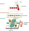
Figure 9.. Schematic of the CTC-iChip.
A schematic of the workflow starting with posts designed for RBC filtration, then inertial focusing of CTCs with removal of WBCs using magnetophoresis. Reprinted by permission from Macmillan Publishers Ltd: Nature Protocols: N. M. Karabacak et al, “Microfluidic, marker-free isolation of CTCs from blood samples,” Nat. Protoc., vol. 9, no. 3, pp. 694–710, copyright year 2014.
Figure 10:
Figure 10:
The multistep immunomagnetic isolation microfluidic set up for capturing specific sub populations of exosomes, lysing to release their surface proteins, and the subsequent immunomagnetic capture of the surface proteins for analysis. Reproduced from M. He, J. Crow, M. Roth, Y. Zeng, and A. K. Godwin, “Integrated immunoisolation and protein analysis of circulating exosomes using microfluidic technology,” Lab Chip, vol. 14, no. 19, p. 3773, 2014 under a “Creative Commons Attribution-Noncommercial 3.0 Unported Licence,” published by the Royal Society of Chemistry [30].
Figure 11:
Figure 11:
The use of photosensitizer-beads acting as a screening assay for immuno-targeted exosomes. Reprinted by permission from Macmillan Publishers Ltd: Y. Yoshioka, N. Kosaka, Y. Konishi, H. Ohta, H. Okamoto, H. Sonoda, R. Nonaka, H. Yamamoto, H. Ishii, M. Mori, K. Furuta, T. Nakajima, H. Hayashi, H. Sugisaki, H. Higashimoto, T. Kato, F. Takeshita, and T. Ochiya, “Ultra-sensitive liquid biopsy of circulating extracellular vesicles using ExoScreen,” Nat. Commun., vol. 5, pp. 1–8, 2014. [93].
Figure 12:
Figure 12:
A schematic of the nanoshearing aided immunoisolation technique for exosome capture. Reprinted with permission from R. Vaidyanathan, M. Naghibosadat, S. Rauf, D. Korbie, L. G. Carrascosa, M. J. A. Shiddiky, and M. Trau, “Detecting Exosomes Speci fi cally: A Multiplexed Device Based on Alternating Current Electrohydrodynamic Induced Nanoshearing,” 2014. Copyright 2014, American Chemical Society[94].
Figure 13:
Figure 13:
Schematic of ciliated micropillars for exosome capture and retention Reproduced from Z. Wang, H.-J. Wu, D. Fine, J. Schmulen, Y. Hu, B. Godin, X. J. Zhang, and X. Liu, “Ciliated micropillars for the microfluidic-based isolation of nanoscale lipid vesicles.” with permission of the Royal Society of Chemistry [42].
Fig 14.
Fig 14.. Principle of operation and SEM image of microfabricated gold electrodes (working electrode and counter electrode).
Reprinted with permission from P. Capaldo, S. R. Alfarano, L. Ianeselli, S. D. Zilio, A. Bosco, P. Parisse, and L. Casalis, “Circulating Disease Biomarker Detection in Complex Matrices: Real-Time, In Situ Measurements of DNA/miRNA Hybridization via Electrochemical Impedance Spectroscopy,” ACS Sensors, vol. 1, no. 8, pp. 1003–1010, 2016. Copyright 2016 American Chemical Society [20].
Figure 15.
Figure 15.. Schematic of gold nanoparticle patterned electrode for electrochemical sensing of miRNAs.
Each subsequent step shows binding of successive molecules for improvement in detection. Reprinted with permission from M. Labib, N. Khan, S. M. Ghobadloo, J. Cheng, J. P. Pezacki, and M. V. Berezovski, “Three-mode electrochemical sensing of ultralow MicroRNA levels,” J. Am. Chem. Soc., vol. 135, no. 8, pp. 3027–3038, 2013. Copyright 2013 American Chemical Society [108].
Figure 16:
Figure 16:. Schematic of components of system with all interconnected PDMS chips, gate microvalves, and the multiplex detection chip.
Reprinted from H. Cai, M. A. Stott, D. Ozcelik, J. W. Parks, A. R. Hawkins, and H. Schmidt, “On-chip wavelength multiplexed detection of cancer DNA biomarkers in blood,” Biomicrofluidics, vol. 10, no. 6, 2016 with the permission of AIP Publishing [110].
Figure 17.
Figure 17.. Components of microfluidic cylindrical illumination for quantitation and sizing of circulating DNA in serum.
Reprinted with permission from K. J. Liu, M. V Brock, I. Shih, and T. Wang, “Decoding Circulating Nucleic Acids in Human Serum Using Microfluidic Single Molecule Spectroscopy,” Am. Chem. Soc., no. 10, pp. 5793–5798, 2010. Copyright 2010 American Chemical Society [118].

References

    1. Tadimety A, Syed A, Nie Y, Long CR, Kready KM, and Zhang JXJ, “Liquid biopsy on chip: a paradigm shift towards the understanding of cancer metastasis,” Integr. Biol, vol. 64, pp. 9–29, 2017. - PubMed
    1. Siegel RL, Miller KD, and Jemal A, “Cancer Statistics, 2017,” vol. 67, no. 1, pp. 7–30, 2017. - PubMed
    1. Hamilton W, “Cancer diagnosis in primary care.,” Br. J. Gen. Pract, vol. 60, no. 571, pp. 121–8, 2010. - PMC - PubMed
    1. Liu C, Pan C, Shen J, Wang H, and Yong L, “MALDI-TOF MS Combined With Magnetic Beads for Detecting Serum Protein Biomarkers and Establishment of Boosting Decision Tree Model for Diagnosis of Colorectal Cancer,” Science (80-.)., vol. 8, no. 1, pp. 39–47, 2011. - PMC - PubMed
    1. Kaur S, Baine MJ, Jain M, Sasson AR, and Batra SK, “Early diagnosis of pancreatic cancer: challenges and new developments,” Biomark. Med, vol. 6, no. 5, pp. 597–612, 2012. - PMC - PubMed

Publication types

Substances

LinkOut - more resources