. 2018 Apr;62(4):255-268.
doi: 10.1111/1348-0421.12579. Epub 2018 Mar 30.

Special AT-rich sequence binding protein 1 is required for maintenance of T cell receptor responsiveness and development of experimental autoimmune encephalomyelitis

Affiliations

Special AT-rich sequence binding protein 1 is required for maintenance of T cell receptor responsiveness and development of experimental autoimmune encephalomyelitis

Yasushi Akiba et al. Microbiol Immunol. 2018 Apr.

Abstract

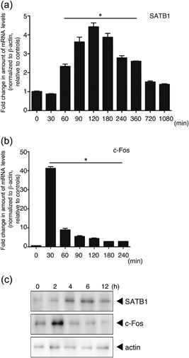
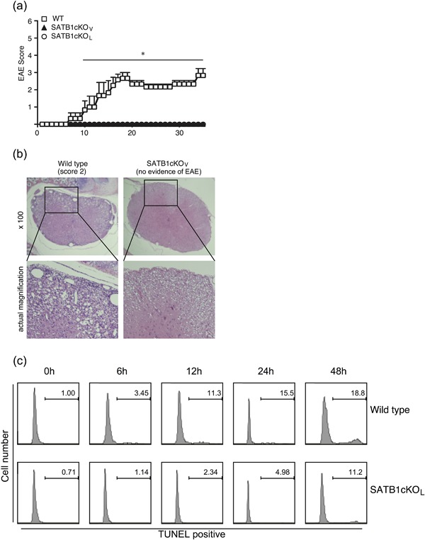
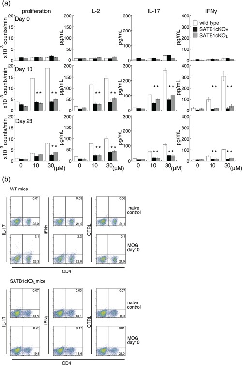
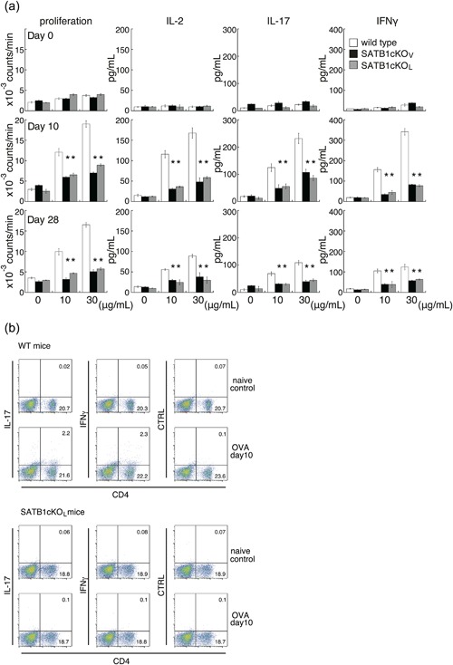
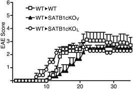
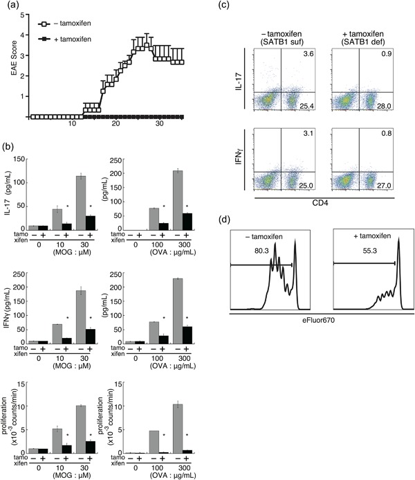
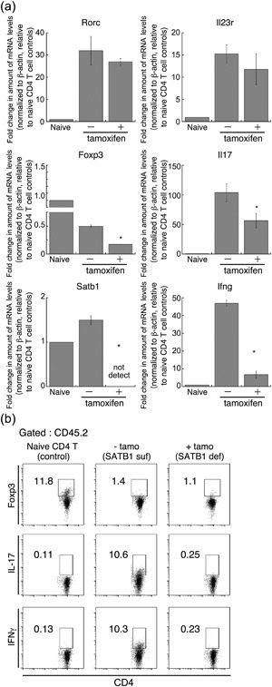
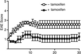
The genome organizer special AT-rich sequence binding protein 1 (SATB1) regulates specific functions through chromatin remodeling in T helper cells. It was recently reported by our team that T cells from SATB1 conditional knockout (SATB1cKO) mice, in which the Satb1 gene is deleted from hematopoietic cells, impair phosphorylation of signaling molecules in response to T cell receptor (TCR) crosslinking. However, in vivo T cell responses upon antigen presentation in the absence of SATB1 remain unclear. In the current study, it was shown that SATB1 modulates T cell antigen responses during the induction and effector phases. Expression of SATB1 was upregulated in response to TCR stimulation, suggesting that SATB1 is important for this antigen response. The role of SATB1 in TCR responses and induced experimental autoimmune encephalomyelitis (EAE) was therefore examined using the myelin oligodendrocyte glycoprotein peptide 35-55 (MOG35-55) and pertussis toxin. SATB1cKO mice were found to be resistant to EAE and had defects in IL-17- and IFN-γ-producing pathogenic T cells. Thus, SATB1 expression appears necessary for T cell function in the induction phase. To examine SATB1 function during the effector phase, a tamoxifen-inducible SATB1 deletion system, SATB1cKO-ER-Cre mice, was used. Encephalitogenic T cells from MOG35-55-immunized SATB1cKO-ER-Cre mice were transferred into healthy mice. Mice that received tamoxifen before the onset of paralysis were resistant to EAE. Furthermore, no disease progression occurred in recipient mice treated with tamoxifen after the onset of EAE. Thus, SATB1 is essential for maintaining TCR responsiveness during the induction and effector phases and may provide a novel therapeutic target for T cell-mediated autoimmune diseases.

Keywords: T cell receptor response; autoimmune disease; experimental autoimmune encephalomyelitis model; special AT-rich sequence binding protein 1.


Figures

Figure 1
Figure 1
Expression of SATB1 in T cells. Naïve CD4 T cells prepared from WT mice were stimulated with anti‐CD3 and anti‐CD28 antibodies for the indicated times. mRNA expression of (a) SATB1 and (b) c‐Fos in these cells was analyzed using real‐time PCR. Transcription of these genes was quantitated relative to β‐actin. *values differ significantly from those in control cells (0 min) under the same conditions (P < 0.05). These results are representative of three independent experiments. (c) Protein expression for SATB1 and c‐Fos, determined by immunoblotting using anti‐SATB1 and anti‐c‐Fos antibodies. Results of one experiment, representative of three independent experiments, is shown.
Figure 2
Figure 2
Failure of SATB1 conditional knockout mice to develop EAE. (a) Mice were immunized s.c. with MOG peptide in CFA, injected i.v. with pertussis toxin on Days 0 and 2, and assessed by clinical scores for 35 days after immunization. * P < 0.05. (b) Photomicrographs of spinal sections stained with hematoxylin and eosin (Day 20) after immunization × 250. Experiments were conducted three times, with essentially similar results. (c) Apoptotic T cell induction by TCR crosslinking was detected by TUNEL staining. Representative flow cytometry plots are shown. [Color figure can be viewed at http://wileyonlinelibrary.com]
Figure 3
Figure 3
In vitro recall response to the MOG peptide for DLN cells from WT, SATB1cKOV, and SATB1cKOL mice. (a) Mice were immunized with MOG in CFA. DLN cells were prepared 10 or 28 days after immunization and incubated with MOG peptide at the indicated doses; proliferation was assessed on Day 3. Production of IL‐2 (Day 1), IL‐17 (Day 3), and IFN‐γ (Day 3) was determined by ELISA. (b) DLN cells were prepared from naïve WT and SATB1cKOL mice or from mice on Day 10 after immunization, as described in the legend of Figure 3(a). DLN cells were incubated with MOG peptide, after which intracellular expression of IL‐17 and IFN‐γ was assessed. The numbers in the light quadrants represent the percentages of all cells. * P < 0.05. One representative result of three independent experiments is shown. [Color figure can be viewed at http://wileyonlinelibrary.com]
Figure 4
Figure 4
In vitro recall response to OVA for DLN cells from WT, SATB1cKOV and SATB1cKOL mice. (a) Mice were immunized with OVA in CFA. DLN cells were prepared 10 or 28 days after immunization and incubated with OVA at the indicated doses; proliferation was assessed on Day 3. Production of IL‐2 (Day 1), IL‐17 (Day 3), and IFN‐γ (Day 3) was determined by ELISA. (b) DLN cells were prepared from naïve WT and SATB1cKOL mice or from mice on Day 10 after immunization, as described in the legend of Figure 4(a). DLN cells were incubated with OVA, after which intracellular expression of IL‐17 and IFN‐γ was assessed. The numbers in the light quadrants represent the percentages of all cells. * P < 0.05. One representative result of three independent experiments is shown. [Color figure can be viewed at http://wileyonlinelibrary.com]
Figure 5
Figure 5
Transfer of encephalitogenic T cells induces EAE in SATB1‐deficient mice. DLN cells were prepared from WT mice 10 days after immunization and incubated with MOG peptide and IL‐23 for 3 days. CD4 T cells were transferred i.v. into naïve WT, SATB1cKOV or SATB1cKOL mice (n = 10). Representative results of three independent experiments are shown as mean EAE scores ± SD.
Figure 6
Figure 6
SATB1 is an essential factor for T cell responses during the effector phase of EAE. (a) Encephalitogenic Th17 cells were prepared from DLN cells derived from SATB1cKOe mice after immunization and stimulated with MOG peptide and IL‐23. CD4 T cells were transferred into naïve WT mice and Satb1 deleted by tamoxifen injection. Clinical status was monitored for 35 days. (b) SATB1cKOe mice were immunized with MOG peptide or OVA in CFA and administered tamoxifen on Days 11 and 13 to delete Satb1. CD4 T cells were prepared from these mice on Day 14 and restimulated with the MOG peptide or OVA presented by irradiated splenocytes from CD45.1 mice. Cytokine production and T cell proliferation were assessed. *, Values differ significantly from those in control cells (SATB1‐sufficient cells) under the same conditions (P < 0.05). (c) IL‐17+ and IFNγ+ CD4 T cells in (b) were analyzed by intracellular staining for cytokines and flow cytometry. (d) CD4 T cells from SATB1cKOe‐OTII mice were labeled with Cell Proliferation Dye eFluor 670 and transferred into RAG2−/− mice. Recipient mice were treated with tamoxifen and then injected with OVA. T cell proliferation was assessed via eFluor 670 dilution in treated (right) versus untreated (left) mice. Plot areas gated on OVA 323‐339 tetramer+ CD4+. [Color figure can be viewed at http://wileyonlinelibrary.com]
Figure 7
Figure 7
Encephalitogenic gene expression in Th17 cells not expressing SATB1. To prepare Th17 cells, DLN cells (CD45.2) from MOG immunized SATB1cKOe mice were skewed using MOG peptide and IL‐23 and then transferred into recipient mice (CD45.1). Some of these mice were treated with tamoxifen on Days 1, 3, and 5 after the transfer. On Day 7, CD4+ CD45.2+ transferred donor T cells were sorted from the spleen and lymph nodes of recipient mice using a FACS Aria III cell sorter. (a) Quantitative PCR was used to measure mRNA expression of Il17A, Ifng, Rorc, Il23r, Satb1 and Foxp3 in donor CD4 T cells. *Values differ significantly from those in control cells (SATB1‐sufficient Th17 cells) under the same conditions (P < 0.05). Experiments were conducted three times, with essentially similar results. (b) CD45.2+ CD4+ Th17 cells from SATB1‐deficient mice (with tamoxifen) or SATB1‐sufficient mice (without tamoxifen), or control CD4 T cells from naïve C57BL/6 mice were assessed for intracellular IL‐17 or Foxp3 expression. The numbers in the squares are the percentages of CD45.2+ CD4+ cells.
Figure 8
Figure 8
Satb1 deletion in Th17 cells after the onset of EAE alleviates clinical evidence of disease in EAE mice. Encephalitogenic Th17 cells were prepared from DLN cells derived from SATB1cKOe mice after immunization and stimulated with MOG peptide and IL‐23. CD4 T cells were transferred into naïve WT mice, and Satb1 deleted by tamoxifen injection after the onset of EAE. Clinical status was monitored for 30 days.

Similar articles

Cited by

References

    1. Phillips J.E., Corces, V.G. (2009) CTCF: Master weaver of the genome. Cell 137: 1194–211. - PMC - PubMed
    1. Schneider R., Grosschedl R (2007) Dynamics and interplay of nuclear architecture, genome organization, and gene expression. Genes Dev 21: 3027–43. - PubMed
    1. Cockerill P.N., Yuen M.H., Garrard W.T. (1987) The enhancer of the immunoglobulin heavy chain locus is flanked by presumptive chromosomal loop anchorage elements. J Biol Chem 262: 5394–97. - PubMed
    1. Zlatanova J.S., van Holde K.E. (1992) Chromatin loops and transcriptional regulation. Crit Rev Eukaryot Gene Exp 2: 211–24. - PubMed
    1. Banan M., Rojas I.C., Lee W.H., King H.L., Harriss J.V., Kobayashi R, Webb C.F., Gottlieb P.D. (1997) Interaction of the nuclear matrix‐associated region (MAR)‐binding proteins, SATB1 and CDP/Cux, with a MAR element (L2a) in an upstream regulatory region of the mouse CD8a gene. J Biol Chem 272: 18440–52. - PubMed

MeSH terms