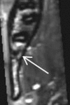
Assessment of impacted and partially impacted lower third molars with panoramic radiography compared to MRI-a proof of principle study
- PMID: 29388826
- PMCID: PMC5991760
- DOI: 10.1259/dmfr.20170371
Assessment of impacted and partially impacted lower third molars with panoramic radiography compared to MRI-a proof of principle study
Abstract
Objectives: Third molars often require surgical removal. Since three-dimensional radiological assessment is often indicated in difficult cases to avoid surgical complications, the radiation burden has to be considered. Here, MRI may offer a dose-free alternative to conventional X-ray techniques. The aim of this retrospective analysis was to evaluate the assessment quality of MRI compared to panoramic radiography in impacted and partially impacted lower third molars.
Methods: Panoramic radiographs and MRI scans of 28 Caucasian patients were assessed twice by four investigators. Wisdom teeth were classified according to Juodzbalys and Daugela 2013.
Results: When radiological lower third molar assessments with panoramic radiography and MRI were compared, staging concurred in 73% in the first round of assessments and 77% in the second.
Conclusions: The presented study demonstrates that MRI not only provides much the same information that panoramic radiography usually does, but also has the advantages of a dose-free three-dimensional view. This may facilitate and shorten third molar surgery. Image interpretation, however, can differ depending on training and experience.
Figures


Similar articles
-
A comparative evaluation of film and digital panoramic radiographs in the assessment of position and morphology of impacted mandibular third molars.Indian J Dent Res. 2011 Mar-Apr;22(2):219-24. doi: 10.4103/0970-9290.84290. Indian J Dent Res. 2011. PMID: 21891889
-
Diagnostic predictability of digital versus conventional panoramic radiographs in the presurgical evaluation of impacted mandibular third molars.Int J Oral Maxillofac Surg. 2009 Nov;38(11):1184-7. doi: 10.1016/j.ijom.2009.06.023. Epub 2009 Aug 5. Int J Oral Maxillofac Surg. 2009. PMID: 19660912
-
The use of cone beam CT for the removal of wisdom teeth changes the surgical approach compared with panoramic radiography: a pilot study.Int J Oral Maxillofac Surg. 2011 Aug;40(8):834-9. doi: 10.1016/j.ijom.2011.02.032. Epub 2011 Apr 19. Int J Oral Maxillofac Surg. 2011. PMID: 21507612
-
Preoperative imaging procedures for lower wisdom teeth removal.Clin Oral Investig. 2008 Dec;12(4):291-302. doi: 10.1007/s00784-008-0200-1. Epub 2008 Apr 30. Clin Oral Investig. 2008. PMID: 18446390 Review.
-
Bilateral maxillary and mandibular fourth molars: a case report and literature review.J Investig Clin Dent. 2011 Nov;2(4):296-9. doi: 10.1111/j.2041-1626.2011.00075.x. Epub 2011 Jun 24. J Investig Clin Dent. 2011. PMID: 25426903 Review.
Cited by
-
Dental MRI-only a future vision or standard of care? A literature review on current indications and applications of MRI in dentistry.Dentomaxillofac Radiol. 2023 Apr;52(4):20220333. doi: 10.1259/dmfr.20220333. Epub 2023 Mar 29. Dentomaxillofac Radiol. 2023. PMID: 36988090 Free PMC article. Review.
-
Low-Field MRI for Dental Imaging in Pediatric Patients With Supernumerary and Ectopic Teeth: A Comparative Study of 0.55 T and Ultra-Low-Dose CT.Invest Radiol. 2025 May 1;60(5):299-310. doi: 10.1097/RLI.0000000000001129. Epub 2024 Oct 23. Invest Radiol. 2025. PMID: 39442499 Free PMC article.
-
Neurosensory Deficits of the Mandibular Nerve Following Extraction of Impacted Lower Third Molars-A Retrospective Study.J Clin Med. 2023 Dec 13;12(24):7661. doi: 10.3390/jcm12247661. J Clin Med. 2023. PMID: 38137730 Free PMC article.
-
Reliability of Orthopantamogram in Lower Third Molar Surgery: Inter- and Intra-observer Agreement.J Pharm Bioallied Sci. 2020 Aug;12(Suppl 1):S190-S193. doi: 10.4103/jpbs.JPBS_57_20. Epub 2020 Aug 28. J Pharm Bioallied Sci. 2020. PMID: 33149454 Free PMC article.
-
Comparison of Preoperative Cone-Beam Computed Tomography and 3D-Double Echo Steady-State MRI in Third Molar Surgery.J Clin Med. 2021 Oct 18;10(20):4768. doi: 10.3390/jcm10204768. J Clin Med. 2021. PMID: 34682896 Free PMC article.
References
-
- Steed MB. The indications for third-molar extractions. J Am Dent Assoc 2014; 145: 570–3. doi: https://doi.org/10.14219/jada.2014.18 - DOI - PubMed
-
- Cassetta M, Pranno N, Barchetti F, Sorrentino V, Lo Mele L. 3.0 Tesla MRI in the early evaluation of inferior alveolar nerve neurological complications after mandibular third molar extraction: a prospective study. Dentomaxillofac Radiol 2014; 43: 20140152. doi: 10.1259/dmfr.20140152 - DOI - PMC - PubMed
Publication types
MeSH terms
LinkOut - more resources
Full Text Sources
Other Literature Sources
Medical

