Review
. 2018 Feb 1;5(1):16.
doi: 10.3390/medicines5010016.

Ovarian Cancers: Genetic Abnormalities, Tumor Heterogeneity and Progression, Clonal Evolution and Cancer Stem Cells

Affiliations
Review

Ovarian Cancers: Genetic Abnormalities, Tumor Heterogeneity and Progression, Clonal Evolution and Cancer Stem Cells

Ugo Testa et al. Medicines (Basel). .

Abstract

Four main histological subtypes of ovarian cancer exist: serous (the most frequent), endometrioid, mucinous and clear cell; in each subtype, low and high grade. The large majority of ovarian cancers are diagnosed as high-grade serous ovarian cancers (HGS-OvCas). TP53 is the most frequently mutated gene in HGS-OvCas; about 50% of these tumors displayed defective homologous recombination due to germline and somatic BRCA mutations, epigenetic inactivation of BRCA and abnormalities of DNA repair genes; somatic copy number alterations are frequent in these tumors and some of them are associated with prognosis; defective NOTCH, RAS/MEK, PI3K and FOXM1 pathway signaling is frequent. Other histological subtypes were characterized by a different mutational spectrum: LGS-OvCas have increased frequency of BRAF and RAS mutations; mucinous cancers have mutation in ARID1A, PIK3CA, PTEN, CTNNB1 and RAS. Intensive research was focused to characterize ovarian cancer stem cells, based on positivity for some markers, including CD133, CD44, CD117, CD24, EpCAM, LY6A, ALDH1. Ovarian cancer cells have an intrinsic plasticity, thus explaining that in a single tumor more than one cell subpopulation, may exhibit tumor-initiating capacity. The improvements in our understanding of the molecular and cellular basis of ovarian cancers should lead to more efficacious treatments.

Keywords: cancer stem cells; chemoresistance; clonal evolution; genetic alterations; metastases; ovarian cancer.


Conflict of interest statement

The authors declare no conflict of interest.

Figures
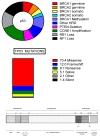
Figure 1
Figure 1
(Top Panel): Mutational spectrum of high-grade serous ovarian cancer (HGS-OvCa). In the figure are reported some of the recurrent genetic abnormalities observed in HGSOC. In the middle of the circle of the figure, TP53 mutations are indicated, occurring in virtually 100% of patients; (Middle Panel): Different types of TP53 mutations and their percentages in HGS-OvCa; (Bottom Panel): Structure of TP53 protein: the different structural and functional domains of the protein are reported. TAD1 and TAD2: Tans Activation Domains 1 and 2; NLS: Nuclear Localization Signal; NES: Nuclear Esportation Signal.
Figure 2
Figure 2
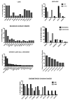
(Top Panel, left): Mutational spectrum observed in LGSOC patients; (Top Panel, right): comparative analysis of the frequency of the main mutations observed through paired comparison odf serous borderline tumors (SBT) and LGSOCs. The data reported in these two figures are from ref. [103]; (Middle Panel, Top, left): Recurrent mutations observed in mucinous cancers of the ovary; (Middle Panel, Top, right): comparative analysis of some recurrently mutated genes at the level of borderline precursor lesions and mucinous carcinomas; (Middle Panel, Bottom, left): Recurrent mutations observed in ovarian clear-cell carcinoma; (Middle Panel, Bottom, right): Recurrent copy number alterations observed in ovarian clear-cell carcinoma. The data shown in these two figures are reported in the references [117,118]; (Bottom Panel): Recurrent gene mutations observed in endometrioid ovarian cancers subdivided into microsatellite instable (MSI) and microsatellite stable (MSS) subgroups. The data shown in this figure are reported from ref. [27].

Similar articles

Cited by

References

    1. Jamal A., Siegel R., Xu J., Ward E. Cancer statistics. CA Cancer J. Clin. 2010;60:277–300. - PubMed
    1. Shih I.M., Kurman R.J. Ovarian tumorigenesis: A proposed model based on morphological and molecular genetic analysis. Am. J. Pathol. 2004;164:1511–1518. doi: 10.1016/S0002-9440(10)63708-X. - DOI - PMC - PubMed
    1. Kurman R.J., Shih I.M. The dualistic model of ovarian carcinogenesis revisited, revised, and expanded. Am. J. Pathol. 2016;186:733–747. doi: 10.1016/j.ajpath.2015.11.011. - DOI - PMC - PubMed
    1. Reid B.M., Permuth J.B., Sellers T.A. Epidemiology of ovarian cancer: Review. Cancer Biol. Med. 2017;14:9–32. - PMC - PubMed
    1. Jones S., Wang T.L., Kurman R.J., Nakayama K., Velculescu V.E., Vogelstein B., Kinzler K.W., Papadopoulos N., Shih I.M. Low-grade serous carcinomas of the ovary contain very few point mutations. J. Pathol. 2012;226:413–420. doi: 10.1002/path.3967. - DOI - PMC - PubMed

LinkOut - more resources