. 2018 Feb 1;13(2):e0192148.
doi: 10.1371/journal.pone.0192148. eCollection 2018.

An oxidized abasic lesion inhibits base excision repair leading to DNA strand breaks in a trinucleotide repeat tract

Affiliations

An oxidized abasic lesion inhibits base excision repair leading to DNA strand breaks in a trinucleotide repeat tract

Jill M Beaver et al. PLoS One. .

Abstract

Oxidative DNA damage and base excision repair (BER) play important roles in modulating trinucleotide repeat (TNR) instability that is associated with human neurodegenerative diseases and cancer. We have reported that BER of base lesions can lead to TNR instability. However, it is unknown if modifications of the sugar in an abasic lesion modulate TNR instability. In this study, we characterized the effects of the oxidized sugar, 5'-(2-phosphoryl-1,4-dioxobutane)(DOB) in CAG repeat tracts on the activities of key BER enzymes, as well as on repeat instability. We found that DOB crosslinked with DNA polymerase β and inhibited its synthesis activity in CAG repeat tracts. Surprisingly, we found that DOB also formed crosslinks with DNA ligase I and inhibited its ligation activity, thereby reducing the efficiency of BER. This subsequently resulted in the accumulation of DNA strand breaks in a CAG repeat tract. Our study provides important new insights into the adverse effects of an oxidized abasic lesion on BER and suggests a potential alternate repair pathway through which an oxidized abasic lesion may modulate TNR instability.


Conflict of interest statement

Competing Interests: The authors have declared that no competing interests exist.

Figures

Fig 1
Fig 1. Generation of DNA substrates containing various types of abasic lesions.
(A) Abasic DNA lesions with modifications to the sugar residue. An oxidized sugar, DOB (left), native sugar, AP (middle), and reduced sugar, THF (right). (B) Formation of single-flap (above) and double-flap (below) intermediates with various types of sugars during BER.
Fig 2
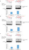
Fig 2. The oxidized abasic lesion DOB inhibits pol β DNA synthesis activity during BER in a CAG repeat duplex.
The effect of the type of abasic lesion on pol β synthesis activity with a substrate containing a single (CAG)2 downstream flap formed during BER of a CAG repeat duplex was examined by measuring pol β synthesis activity on the substrate containing a DOB (left panel), native sugar (middle panel), or THF (right panel). Lanes 1, 7, and 13 indicate the substrate only. Lanes 2–6, lanes 8–12, and lanes 14–18 indicate the substrate incubated with pol β (2.5 nM) for a time course of 2, 5, 10, 15, and 30 minutes. Substrates were 32P-labeled at the 5’-end of the upstream strand and are illustrated above each gel. The experiments were repeated at least three times. Representative gels are shown. The quantification of the data is presented below the gels. Two-way ANOVA with Tukey’s multiple comparison posttests was used to determine statistically significant differences. "*" denotes P < 0.05, compared to the DOB-containing substrates.
Fig 3
Fig 3. The oxidized abasic lesion DOB inhibits pol β DNA synthesis activity during BER in a small CAG hairpin loop.
The effect of the type of abasic lesion on pol β synthesis activity with a short (CAG)3/(CAG)2 double-flap intermediate formed during BER of a (CAG)5 hairpin was examined by measuring pol β synthesis activity on the substrate containing a DOB (left panel), native sugar (middle panel), or THF (right panel). Lanes 1, 7, and 13 indicate the substrate only. Lanes 2–6, lanes 8–12, and lanes 14–18 indicate the substrate incubated with pol β (2.5 nM) for a time course of 2, 5, 10, 15, and 30 minutes. Substrates were 32P-labeled at the 5’-end of the upstream strand and are illustrated above each gel. The experiments were repeated at least three times. Representative gels are shown. The quantification of the data is presented below the gels. Two-way ANOVA with Tukey’s multiple comparison posttests was used to determine statistically significant differences. "*" denotes P < 0.05, compared to the DOB-containing substrates.
Fig 4
Fig 4. Wild-type pol β crosslinks with DOB and AP, whereas pol β K72A mutant only crosslinks with DOB.
The formation of a pol β•DOB crosslink complex (A) and pol β K72A mutant•DOB crosslink complex (B) was measured using the nicked flap substrates and double-flap substrates containing DOB as described in the “Methods”. Lanes 1 and 7 contain the unphotolyzed substrate only. Lanes 2 and 8 represent reaction mixtures with the unphotolyzed substrates and pol β (1 μM). Lanes 3 and 9 indicate reaction mixtures containing the unphotolyzed substrates and pol β (1 μM) in the presence of 100 mM NaBH4. Lanes 4 and 10 indicate the photolyzed substrate only. Lanes 5 and 11 indicate reaction mixtures with the photolyzed substrates and pol β (1 μM). Lanes 6 and 12 indicate reaction mixtures containing the photolyzed substrates and pol β (1 μM) in the presence of 100 mM NaBH4. (C) The formation of pol β•AP crosslink and pol β K72A mutant•AP crosslink was examined using the nicked flap substrates and double-flap substrates containing a 5’-uracil as described in the “Methods”. Lanes 1 and 6 indicate the substrate only. Lanes 2 and 7 indicate reaction mixtures with 1 μM pol β only. Lanes 3 and 8 indicate reaction mixtures containing 1 μM pol β in the presence of 100 mM NaBH4. Lanes 4 and 9 indicate reaction mixtures with 1 μM pol β K72A mutant only. Lanes 5 and 10 indicate reaction mixtures with 1 μM pol β K72A mutant in the presence of 100 mM NaBH4. Substrates were 32P-labeled at the 3’-end of the downstream strand and are illustrated above each gel. Each experiment was done in triplicate.
Fig 5
Fig 5. The oxidized abasic lesion does not significantly affect FEN1 5’-flap cleavage activity during BER in a CAG repeat duplex.
The effect of the pol β-DOB crosslink on FEN1 cleavage activity on a substrate containing a single (CAG)2 downstream flap intermediate formed during BER of CAG repeat duplex was explored by measuring FEN1 cleavage activity on the substrate containing a DOB (left panel), native sugar (middle panel), or THF (right panel). Lanes 1, 5, and 9 indicate substrate only. Lanes 2, 6, and 10 indicate substrate incubated with FEN1 (1 nM) only. Lanes 3, 7, and 8 indicate the substrate pre-incubated with pol β (2.5 nM) for 1 min prior to incubation with FEN1 (1 nM). Lanes 4, 8, and 12 indicate substrates pre-incubated with pol β (2.5 nM) for 5 min prior to incubation with FEN1 (1 nM). Substrates were 32P-labeled at the 3’-end of the upstream strand and are illustrated above each gel. The experiments were repeated at least three times. Representative gels are shown. The quantification of the data is presented below the gels. Two-way ANOVA with Tukey’s multiple comparison posttests was used to determine statistically significant differences. "*" denotes P < 0.05, compared to the DOB-containing substrates.
Fig 6
Fig 6. The oxidized abasic lesion does not affect FEN1 5’-flap cleavage activity during BER in a small CAG hairpin loop.
The effect of the pol β-DOB crosslink on FEN1 cleavage activity on a short (CAG)3/(CAG)2 double-flap intermediate formed during BER of a (CAG)5 hairpin was examined by measuring FEN1 cleavage activity on the substrate containing a DOB (left panel), native sugar (middle panel), or THF (right panel). Lanes 1, 5, and 9 indicate the substrate only. Lanes 2, 6, and 10 indicate substrates incubated with FEN1 (1 nM) only. Lanes 3, 7, and 8 indicate substrates pre-incubated with pol β (5 nM) for 1 min prior to incubation with FEN1 (1 nM). Lanes 4, 8, and 12 indicate substrates pre-incubated with pol β (5 nM) for 5 min prior to incubation with FEN1 (1 nM). Substrates were 32P-labeled at the 3’-end of the upstream strand and are illustrated above each gel. The experiments were repeated at least three times. Representative gels are shown. The quantification of the data is presented below the gels. Two-way ANOVA with Tukey’s multiple comparison posttests was used to determine statistically significant differences. "*" denotes P < 0.05, compared to the DOB-containing substrates.
Fig 7
Fig 7. An oxidized abasic lesion (DOB) decreases BER efficiency in a CAG repeat duplex.
The effect of abasic lesion type on BER efficiency during BER in a CAG repeat duplex was examined by reconstituting BER with a substrate containing a single (CAG)2 downstream flap intermediate formed during BER of a CAG repeat duplex containing a DOB (left panel), native sugar (middle panel), or THF (right panel). Lanes 1, 6, and 11 indicate the substrate only. Lanes 2, 7, and 12 indicate the substrate incubated with FEN1 (1 nM) only. Lanes 3, 8, and 13 indicate reaction mixtures incubated with FEN1 (1 nM) and LIG I (2.5 nM). Lanes 4, 9, and 14 indicate reaction mixtures incubated with FEN1 (1 nM), LIG I (2.5 nM), and pol β (1 nM). Lanes 5, 10, and 15 indicate synthesized size markers as indicated to the left of the gel. Graphs indicating the percentage of repaired products are shown below each gel. Each experiment was done in triplicate, and only the representative gels are shown in the figures. Substrates were 32P-labeled at the 5’-end of the upstream strand and are illustrated above each gel. Percentage of repair product (%) = intensity of repaired product/(intensity of repaired product + intensity of residual substrate)×100. The quantification data are illustrated below the gels. Two-way ANOVA with Tukey’s multiple comparison posttests was used to determine statistically significant differences. "*" denotes P < 0.05, compared to DOB-containing substrates. "#" denotes P < 0.05, compared to repair reactions in the absence of pol β.
Fig 8
Fig 8. An oxidized abasic lesion (DOB) decreases repair efficiency during BER in a small CAG hairpin loop.
The effect of abasic lesion type on BER efficiency during BER in a small CAG hairpin loop was examined by reconstituting BER with the short (CAG)3/(CAG)2 double-flap intermediate formed during BER in a (CAG)5 hairpin containing a DOB (left panel), native sugar (middle panel), or THF (right panel). Lanes 1, 6, and 11 indicate the substrate only. Lanes 2, 7, and 12 indicate the substrate incubated with FEN1 (1 nM) only. Lanes 3, 8, and 13 indicate reaction mixtures incubated with FEN1 (1 nM) and LIG I (2.5 nM). Lanes 4, 9, and 14 indicate reaction mixtures incubated with FEN1 (1 nM), LIG I (2.5 nM), and pol β (1 nM). Lanes 5, 10, and 15 indicate synthesized size markers as indicated to the left of the gel. Graphs indicating the percentage of repaired products are shown below each gel. Each experiment was done in triplicate, and only the representative gels are shown in the figures. Substrates were 32P-labeled at the 5’-end of the upstream strand and are illustrated above each gel. Percentage of repair product (%) = intensity of repaired product/(intensity of repaired product + intensity of residual substrate)×100. The quantification data are illustrated below the gels. Two-way ANOVA with Tukey’s multiple comparison posttests was used to determine statistically significant differences. "*" denotes P < 0.05, compared to the DOB-containing substrates. "#" denotes P < 0.05, compared to repair reactions in the absence of pol β.
Fig 9
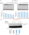
Fig 9. An oxidized abasic lesion inhibits LIG I activity by a DOB-LIG I crosslink.
(A) The effect of an oxidized lesion on LIG I activity was examined by incubating the nicked DNA substrate with LIG I in the presence of increasing concentrations of DOB—(left panel) or THF—(right panel) containing DNA (0–1000 nM). Lanes 1 and 9 indicate the substrate alone. Lanes 2 and 10 indicate the substrate incubated with 2.5 nM LIG I only. Lanes 3–8 and lanes 11–16 indicate the substrate incubated with 2.5 nM LIG I and increasing concentrations of lesion-containing DNA (0–1000 nM), as indicated. Graphs indicating the percentage of ligated products are shown below each gel. Substrates were 32P-labeled at the 5’-end of the upstream strand and are illustrated above each gel. Two-way ANOVA with Tukey’s multiple comparison posttests was used to determine statistically significant differences. "*" denotes P < 0.05, compared to the DOB-containing substrates. (B) The formation of a LIG I•DOB crosslink complex was determined as described in the “Methods”. Lane 1 indicates the unphotolyzed substrate only. Lane 2 indicates reaction mixtures with the unphotolyzed substrates and LIG I (1 μM). Lanes 3–5 indicate reaction mixtures containing the unphotolyzed substrates and LIG I (1 μM) in the presence of 20, 50, and 100 mM NaBH4, respectively. Lane 6 indicates the photolyzed substrate only. Lane 7 indicates the reaction mixtures with photolyzed substrates and LIG I (1 μM). Lanes 8–10 indicate reaction mixtures containing the photolyzed substrates and LIG I (1 μM) in the presence of 20, 50, and 100 mM NaBH4, respectively. Each experiment was done in triplicate, and only representative gels are shown in the figures.

Similar articles

Cited by

References

    1. Paulson HL, Fischbeck KH. Trinucleotide repeats in neurogenetic disorders. Annu Rev Neurosci. 1996;19:79–107. doi: 10.1146/annurev.ne.19.030196.000455 - DOI - PubMed
    1. McMurray CT. Mechanisms of trinucleotide repeat instability during human development. Nat Rev Genet. 2010;11(11):786–99. doi: 10.1038/nrg2828 - DOI - PMC - PubMed
    1. Wells RD, Dere R, Hebert ML, Napierala M, Son LS. Advances in mechanisms of genetic instability related to hereditary neurological diseases. Nucleic Acids Res. 2005;33(12):3785–98. doi: 10.1093/nar/gki697 - DOI - PMC - PubMed
    1. Usdin K, House NC, Freudenreich CH. Repeat instability during DNA repair: Insights from model systems. Crit Rev Biochem Mol Biol. 2015;50(2):142–67. doi: 10.3109/10409238.2014.999192 - DOI - PMC - PubMed
    1. Wang G, Vasquez KM. Impact of alternative DNA structures on DNA damage, DNA repair, and genetic instability. DNA Repair (Amst). 2014;19:143–51. - PMC - PubMed

Publication types

MeSH terms

Substances