. 2018 Feb 1;13(2):e0191802.
doi: 10.1371/journal.pone.0191802. eCollection 2018.

Rhodopsin optogenetic toolbox v2.0 for light-sensitive excitation and inhibition in Caenorhabditis elegans

Affiliations

Rhodopsin optogenetic toolbox v2.0 for light-sensitive excitation and inhibition in Caenorhabditis elegans

Amelie Bergs et al. PLoS One. .

Abstract

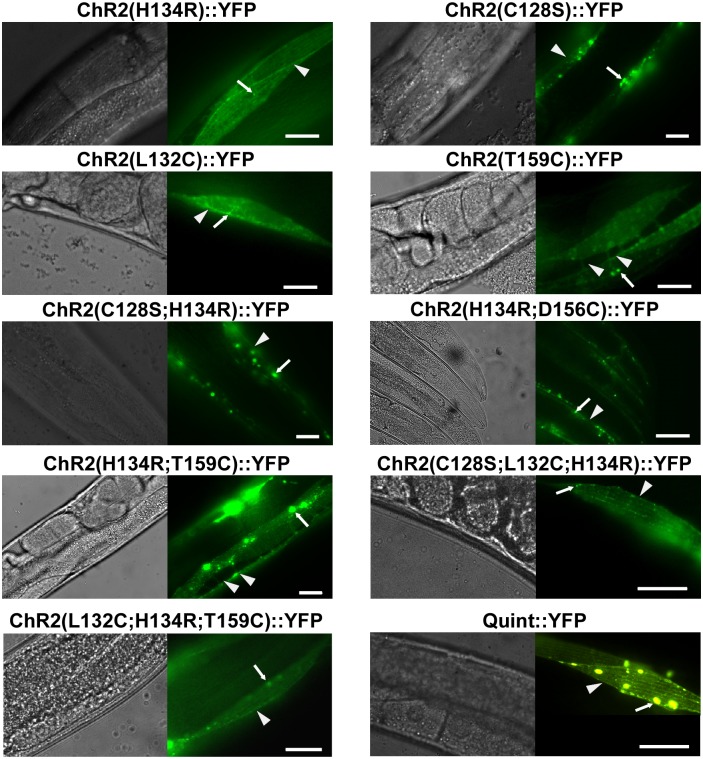
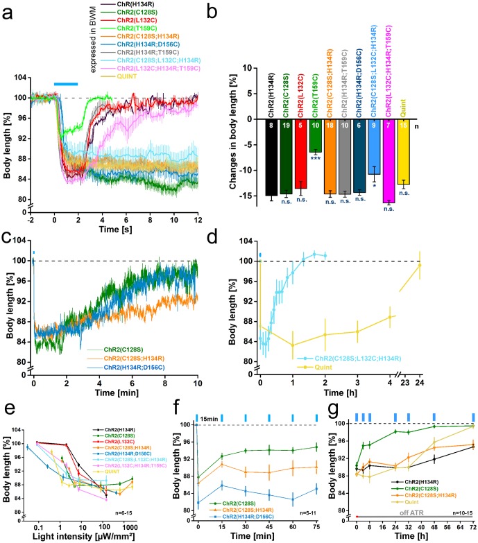
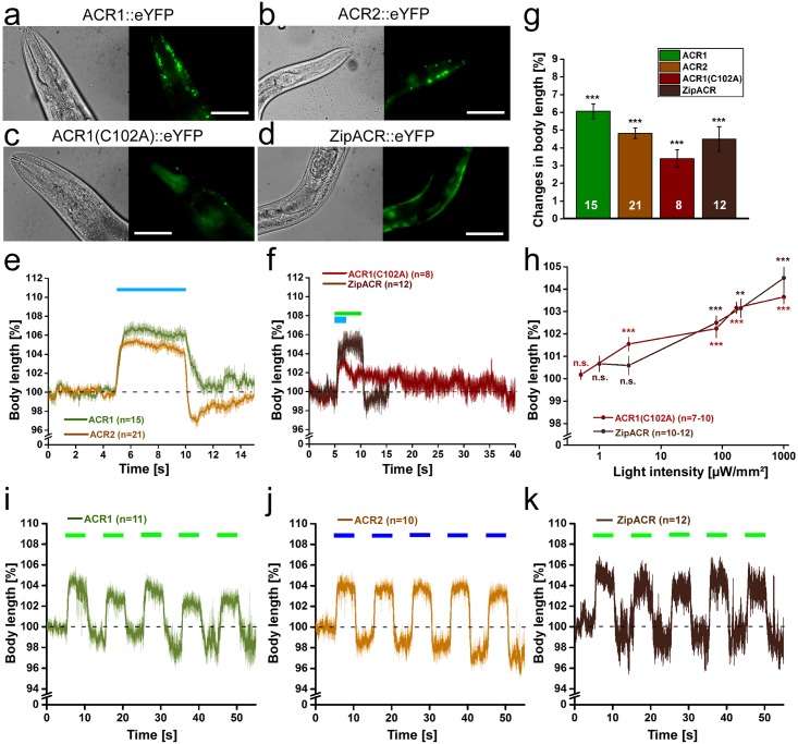
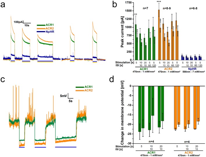
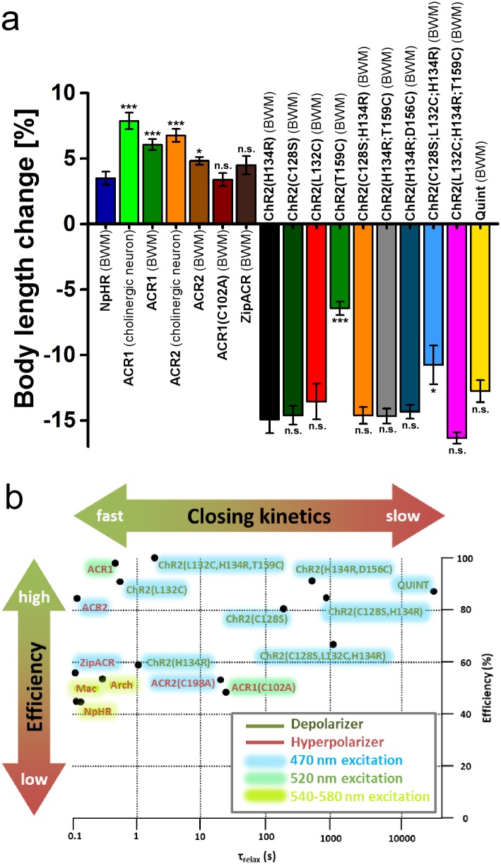
In optogenetics, rhodopsins were established as light-driven tools to manipulate neuronal activity. However, during long-term photostimulation using channelrhodopsin (ChR), desensitization can reduce effects. Furthermore, requirement for continuous presence of the chromophore all-trans retinal (ATR) in model systems lacking sufficient endogenous concentrations limits its applicability. We tested known, and engineered and characterized new variants of de- and hyperpolarizing rhodopsins in Caenorhabditis elegans. ChR2 variants combined previously described point mutations that may synergize to enable prolonged stimulation. Following brief light pulses ChR2(C128S;H134R) induced muscle activation for minutes or even for hours ('Quint': ChR2(C128S;L132C;H134R;D156A;T159C)), thus featuring longer open state lifetime than previously described variants. Furthermore, stability after ATR removal was increased compared to the step-function opsin ChR2(C128S). The double mutants C128S;H134R and H134R;D156C enabled increased effects during repetitive stimulation. We also tested new hyperpolarizers (ACR1, ACR2, ACR1(C102A), ZipACR). Particularly ACR1 and ACR2 showed strong effects in behavioral assays and very large currents with fast kinetics. In sum, we introduce highly light-sensitive optogenetic tools, bypassing previous shortcomings, and thus constituting new tools that feature high effectiveness and fast kinetics, allowing better repetitive stimulation or investigating prolonged neuronal activity states in C. elegans and, possibly, other systems.


Conflict of interest statement

Competing Interests: The authors have declared that no competing interests exist.

Figures

Fig 1
Fig 1. ChR2 variants expressed in body-wall muscle cells localize to membranes.
DIC images (left panels) and fluorescence micrographs (right panels) showing expression of ChR2(H134R)::YFP, ChR2(C128S)::YFP, ChR2(L132C)::YFP, ChR2(T159C)::YFP ChR2(C128S;H134R)::YFP, ChR2(H134R;D156C)::YFP, ChR2(H134R;T159C)::YFP, ChR2(C128S;L132C;H134R)::YFP, ChR2(L132C;H134R;T159C)::YFP, and Quint::YFP in body-wall muscle cells of C. elegans, as indicated. Arrows mark aggregates, arrowheads mark plasma membrane. Scale bar is 10 μm.
Fig 2
Fig 2. ChR2 variants expressed in body-wall muscle cells enable prolonged depolarization and body contractions.
(a) Body length of animals expressing ChR2(H134R)::YFP, ChR2(C128S)::YFP, ChR2(L132C)::YFP, ChR2(T159C)::YFP ChR2(C128S;H134R)::YFP, ChR2(H134R;D156C)::YFP, ChR2(H134R;T159C)::YFP, ChR2(C128S;L132C;H134R)::YFP, ChR2(L132C;H134R;T159C):.YFP, and Quint::YFP in body-wall muscle cells of C. elegans during and after a blue light stimulus (0.2 mW/mm2; 1 or 2 s; 450–490 nm; n = 5–19). b) Maximal changes in body length of all depolarizers tested. Shown is the mean normalized change in body length (± SEM) relative to the initial length of the animal. Significance given refers to ChR2(H134R): *p<0.05, ***p<0.001. (c) Same as in a) but displayed over 600 s for ChR2(C128S)::YFP, ChR2(C128S;H134R)::YFP, and ChR2(H134R;D156C)::YFP (n = 7–19). (d) Body length of animals expressing ChR2(C128S;L132C;H134R) (n = 5–6; 2 s illumination; 470 nm; 1 mW/mm2) or Quint (n = 8–14; 5 s illumination; 450–490 nm; 2.4 mW/mm2) up to 24h after a blue light stimulus (450–490 nm). (e) Dependence of body contractions on light intensity in strains listed in (a). Reductions in body length were recorded in response to 1, 2 or 5 s light stimuli (450–490 nm) of light intensities in the range of 0.03–2.41 mW/mm2 (n = 6–15). (f) Repeated stimulation of animals expressing ChR2(C128S)::YFP, ChR2(C128S;H134R)::YFP, and ChR2(H134R;D156C)::YFP. 2 s blue light pulses (0.2 mW/mm2 ((C128S), (C128S;H134R)) or 1 mW/mm2 (H134R;D156C); 450–490 nm; n = 5–11) were presented with ISI of 15 min. (g) Off-ATR excitability of animals up to 72 h after placing them on fresh NGM plates in absence of ATR (5s; 2.4 mW/mm2; 450–490 nm; n = 10–15). Shown is the mean normalized body length (± SEM) calculated relative to the initial length of the animal; n = number of animals. Blue bar indicates illumination period.
Fig 3
Fig 3. ACRs mediate strong hyperpolarization and body elongation.
Expression of ACR1::eYFP (a), ACR2::eYFP (b), as well as ACR1(C102A)::eYFP (c) and ZipACR::eYFP (d) in body-wall muscle cells of C. elegans. Scale bar is 50 μm. (e) Body length of animals expressing ACR1 or ACR2 during and after a 5 s light stimulus (1 mW/mm2; 470 nm). Shown is the mean normalized body length (± SEM) relative to the initial length of the animal. (f) Body length during and after a light stimulus (1 mW/mm2) of animals expressing ACR1(C102A) (470 nm, 2 s) or ZipACR (520 nm, 5 s). (g) Maximal changes in body length induced by the tested hyperpolarizers. Shown is the mean normalized change in body length (± SEM) relative to the initial length of the animal. (h) Light intensity dependence of the body elongation of animals expressing ACR1(C102A) or ZipACR in body-wall muscle cells. (i) Body length in response to repetitive photostimulation (5 s, 5 s ISI, 80 μW/mm2, 470 or 520 nm) of ACR1, ACR2 (j) or ZipACR (k) in animals expressing the respective channel in body-wall muscles; n = number of animals. Blue and green bars indicate illumination period. *p<0.05, **p<0.01, ***p<0.001.
Fig 4
Fig 4. ACR1 and ACR2 mediate large photocurrents and hyperpolarization in patch-clamped BWMs.
(a) Example current traces of body-wall muscle cells expressing ACR1, ACR2, or NpHR during 5 s, 10 s, and 20 s photostimulation (blue or yellow light, as indicated by bars) with 10 s ISIs. (b) Group data of experiments described in a). The paired bars at each time point represent peak currents (1st bar) and plateau currents (2nd bar). Significance refers to NpHR. (c) As in a), but showing voltage traces, before, during and after 5 s, 10 s, and 20 s photostimulation with 10 s ISI. (d) Group data for experiments shown in c). The paired bars at each time point represent peak (1st bar) and plateau (2nd bar) changes in the membrane potential. ACRs were illuminated at 470 nm (1 mW/mm2), NpHR at 590 nm (7 mW/mm2); n = number of animals. Displayed are mean ± SEM of currents or voltage in b) and c). **p<0.01, ***p<0.001.
Fig 5
Fig 5. Evaluation of de- and hyperpolarizers characterized in this paper.
a) Changes in body length induced by de- and hyperpolarizers. Shown is the mean normalized change in body length (± SEM) relative to the initial length of the animal; n = number of animals. Statistical differences for hyperpolarizers are referring to NpHR, statistical differences for depolarizers are referring to ChR2(H134R). b) Scheme of optogenetic de- and hyperpolarizers expressed in BWMs of C. elegans classified by closing kinetics (τrelax) and efficiency. The efficiency was determined as follows: Depolarizers—Relative comparison of contractions, induced by the respective tool, at 200 μW/mm2 to the maximum possible contraction; Hyperpolarizers—Relative comparison of relaxations, induced by the respective tool, at 1 mW/mm2 to the maximum possible relaxation. Hence, efficiency not only refers to the maximum possible changes in body length upon light saturation, but also includes information about the tool’s light sensitivity. Therefore, some tools receive lower efficiencies, though they exhibit comparable maximum effects. Color shades indicate the light color of the respective tool’s excitation wavelength.

Similar articles

Cited by

References

    1. Knöpfel T, Lin MZ, Levskaya A, Tian L, Lin JY, Boyden ES. Toward the second generation of optogenetic tools. J Neurosci. 2010;30(45):14998–5004. doi: 10.1523/JNEUROSCI.4190-10.2010 - DOI - PMC - PubMed
    1. Yizhar O, Fenno LE, Davidson TJ, Mogri M, Deisseroth K. Optogenetics in neural systems. Neuron. 2011;71(1):9–34. doi: 10.1016/j.neuron.2011.06.004 - DOI - PubMed
    1. Deisseroth K. Optogenetics: 10 years of microbial opsins in neuroscience. Nat Neurosci. 2015;18(9):1213–25. doi: 10.1038/nn.4091 - DOI - PMC - PubMed
    1. Kocabas A, Shen CH, Guo ZV, Ramanathan S. Controlling interneuron activity in Caenorhabditis elegans to evoke chemotactic behaviour. Nature. 2012;490(7419):273–7. doi: 10.1038/nature11431 - DOI - PMC - PubMed
    1. Erbguth K, Prigge M, Schneider F, Hegemann P, Gottschalk A. Bimodal activation of different neuron classes with the spectrally red-shifted channelrhodopsin chimera C1V1 in Caenorhabditis elegans. PLoS ONE. 2012;7(10):e46827 doi: 10.1371/journal.pone.0046827 - DOI - PMC - PubMed

Publication types