. 2018 Feb 1;13(2):e0192352.
doi: 10.1371/journal.pone.0192352. eCollection 2018.

Serum amyloid A3 is required for normal weight and immunometabolic function in mice

Affiliations

Serum amyloid A3 is required for normal weight and immunometabolic function in mice

Jennifer L Ather et al. PLoS One. .

Abstract

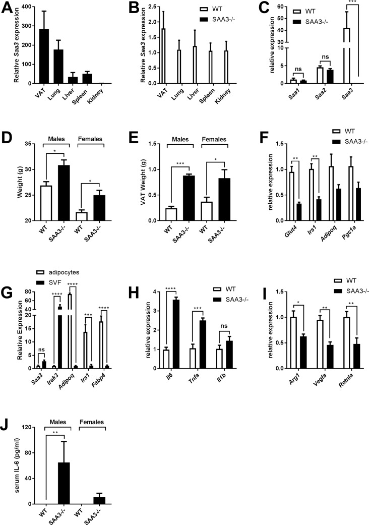
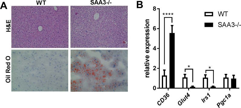
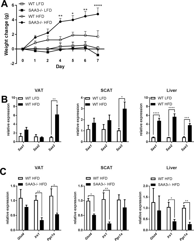
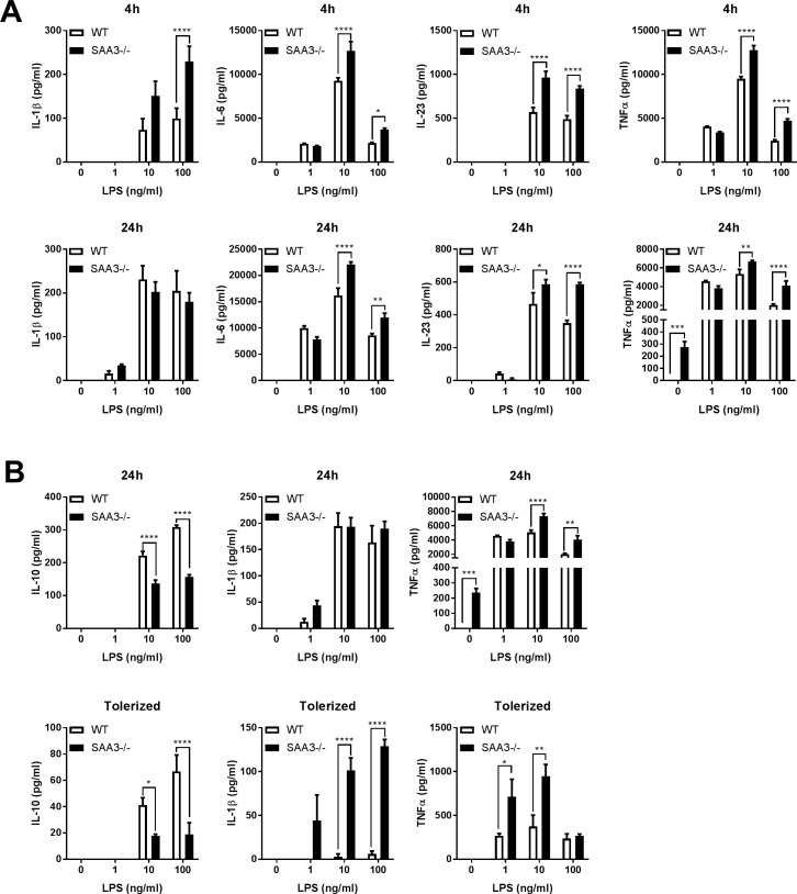
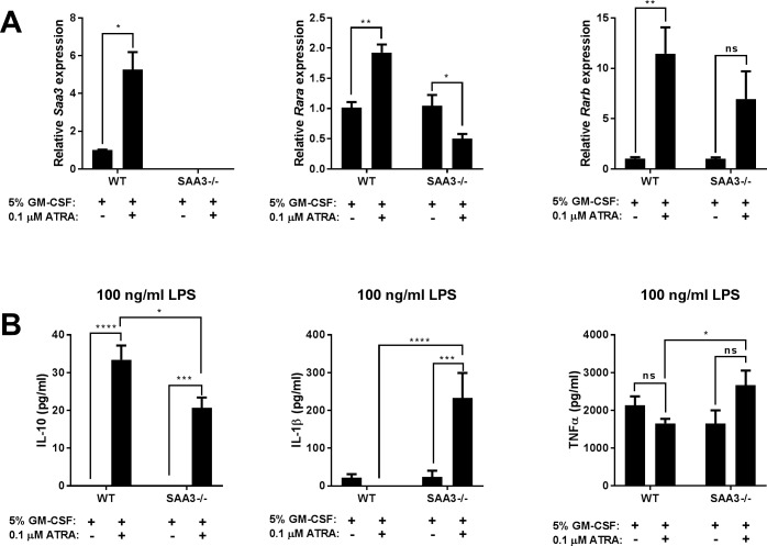
Serum amyloid A (SAA) is an apolipoprotein that is robustly upregulated in numerous inflammatory diseases and has been implicated as a candidate pro-inflammatory mediator. However, studies comparing endogenous SAAs and recombinant forms of the acute phase protein have generated conflicting data on the function of SAA in immunity. We generated SAA3 knockout mice to evaluate the contribution of SAA3 to immune-mediated disease, and found that mice lacking SAA3 develop adult-onset obesity and metabolic dysfunction along with defects in innate immune development. Mice that lack SAA3 gain more weight, exhibit increased visceral adipose deposition, and develop hepatic steatosis compared to wild-type littermates. Leukocytes from the adipose tissue of SAA3-/- mice express a pro-inflammatory phenotype, and bone marrow derived dendritic cells from mice lacking SAA3 secrete increased levels of IL-1β, IL-6, IL-23, and TNFα in response to LPS compared to cells from wild-type mice. Finally, BMDC lacking SAA3 demonstrate an impaired endotoxin tolerance response and inhibited responses to retinoic acid. Our findings indicate that endogenous SAA3 modulates metabolic and immune homeostasis.


Conflict of interest statement

Competing Interests: The authors have declared that no competing interests exist.

Figures

Fig 1
Fig 1. Characterization of SAA3-/- mice.
Levels of basal Saa3 expression from wild-type (WT) mice were measured from VAT, lung, liver, spleen, and kidney. Relative levels were compared to the kidney (A). WT and SAA3 knockout (SAA3-/-) littermates were analyzed by Q-PCR for Saa3 gene expression in the same organs to confirm the deletion of Saa3 in the SAA3-/- mice (B). Characterization of different SAA isoforms was performed by Q-PCR from VAT (C). WT and SAA3-/- mice were analyzed at 18 weeks of age for total body weight (D) and VAT weight (E). Insulin sensitive gene expression was analyzed by Q-PCR from VAT (F). Adipocyte and stromal vascular fractions were confirmed for cell-specific genes in wild-type mice (G). SVF from the VAT of WT and SAA3-/- mice were analyzed by Q-PCR for pro-inflammatory genes (H) and M2-related genes (I). Serum from WT and SAA3-/- mice was analyzed for circulating cytokines (J) n = 3-9/group. * = p<0.05, ** = p<0.01, *** = p<0.005, **** = p<0.001.
Fig 2
Fig 2. Livers from SAA3-/- mice demonstrate early signs of non-alcoholic fatty liver disease.
Liver tissue from WT and SAA3-/- mice at 18 weeks of age were fixed in 10% neutral-buffered formalin and stained for H&E (A, top row) and Oil-Red O (A, bottom row). Representative images are presented. Q-PCR analysis from liver was performed for genes that regulate insulin sensitivity (B). n = 3-4/group. *p = <0.05, **** = p<0.001.
Fig 3
Fig 3. High fat diet leads to more substantial weight gain and insulin-sensitive gene repression in SAA3-/- mice.
18 week old wild type (WT) and SAA3-/- mice were placed on high-fat diet (HFD = 60% kCal from fat) or low-fat diet (LFD = 10% kCal from fat) for 7 days. Total body weight was measured each day (A). Comparisons displayed are between HFD-fed WT and SAA3-/- mice. The expression of SAA isoforms in VAT was analyzed from WT mice on the LFD and HFD (B). Insulin-sensitive gene expression was analyzed by Q-PCR from VAT, subcutaneous adipose tissue (SCAT), or liver of WT and SAA3-/- mice fed HFD (C). n = 4-5/group. * = p<0.05, ** = p<0.01, *** = p<0.005, **** = p<0.001.
Fig 4
Fig 4. SAA3-/- mice display altered myeloid cell responses.
Bone marrow derived dendritic cells generated from WT and SAA3-/- mice were challenged with increasing doses of LPS for 4 and 24 hours. IL-1β, IL-6, IL-23, and TNFα were measured from cell supernatants by ELISA (A). BMDC were challenged for 24 hours with increasing doses of LPS (24h). These cells were then restimulated with the same dose of LPS for a further 24 hours (Tolerized) and supernatants were analyzed for IL-1β, IL-10, and TNFα by ELISA (B). n = 3-7/group. * = p<0.05, ** = p<0.01, *** = p<0.005, **** = p<0.001.
Fig 5
Fig 5. Lack of SAA3 inhibits dendritic cell responses to retinoic acid.
Bone marrow derived cells were isolated from WT and SAA3-/- mice and differentiated into dendritic cells for 7 days in the presence of 5% GM-CSF or 5% GM-CSF + 0.1 μM all-trans-retinoic acid (ATRA). At day 7, cells were analyzed for Saa3, Rara, and Rarb gene expression by Q-PCR (A). At day 7, cells were also challenged with 100 ng/ml LPS for 24 hours, and IL-10, IL-1β, and TNFα were measured from cell supernatants by ELISA (B). n = 3-7/group. * = p<0.05, ** = p<0.01, *** = p<0.005, **** = p<0.001.

References

    1. Uhlar CM, Whitehead AS. Serum amyloid A, the major vertebrate acute-phase reactant. Eur J Biochem. 1999;265(2):501–23. . - PubMed
    1. Steel DM, Sellar GC, Uhlar CM, Simon S, DeBeer FC, Whitehead AS. A constitutively expressed serum amyloid A protein gene (SAA4) is closely linked to, and shares structural similarities with, an acute-phase serum amyloid A protein gene (SAA2). Genomics. 1993;16(2):447–54. doi: 10.1006/geno.1993.1209 . - DOI - PubMed
    1. Ramadori G, Sipe JD, Colten HR. Expression and regulation of the murine serum amyloid A (SAA) gene in extrahepatic sites. J Immunol. 1985;135(6):3645–7. . - PubMed
    1. Ather JL, Ckless K, Martin R, Foley KL, Suratt BT, Boyson JE, et al. Serum amyloid A activates the NLRP3 inflammasome and promotes Th17 allergic asthma in mice. J Immunol. 2011;187(1):64–73. doi: 10.4049/jimmunol.1100500 ; PubMed Central PMCID: PMC3119761. - DOI - PMC - PubMed
    1. Derebe MG, Zlatkov CM, Gattu S, Ruhn KA, Vaishnava S, Diehl GE, et al. Serum amyloid A is a retinol binding protein that transports retinol during bacterial infection. Elife. 2014;3:e03206 doi: 10.7554/eLife.03206 ; PubMed Central PMCID: PMC4129439. - DOI - PMC - PubMed

Publication types