. 2018 Feb 28;46(4):1565-1583.
doi: 10.1093/nar/gky068.

Metabolic and chemical regulation of tRNA modification associated with taurine deficiency and human disease

Affiliations

Metabolic and chemical regulation of tRNA modification associated with taurine deficiency and human disease

Kana Asano et al. Nucleic Acids Res. .

Abstract

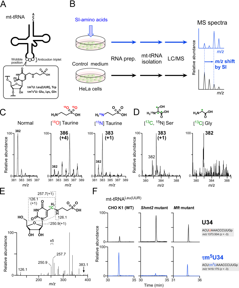
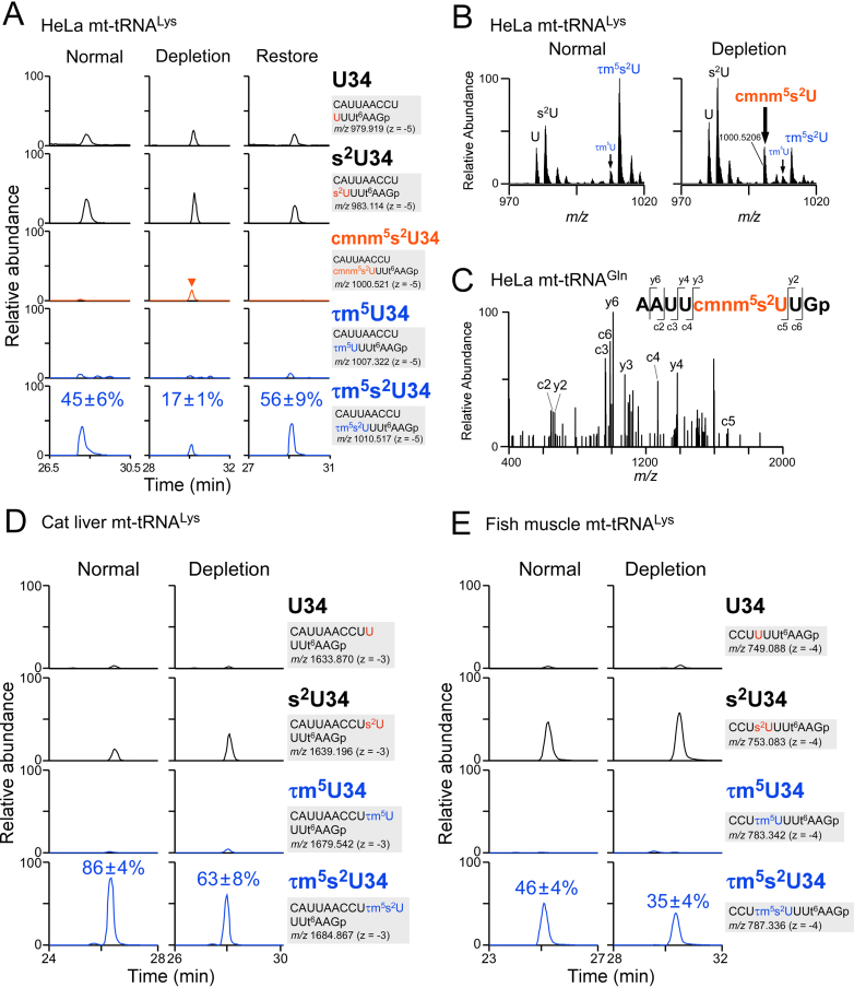
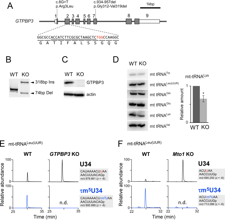
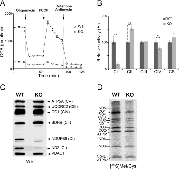
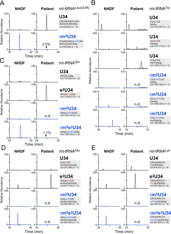
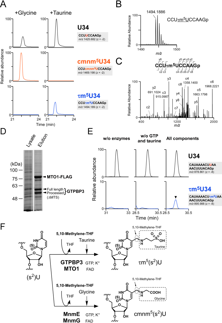
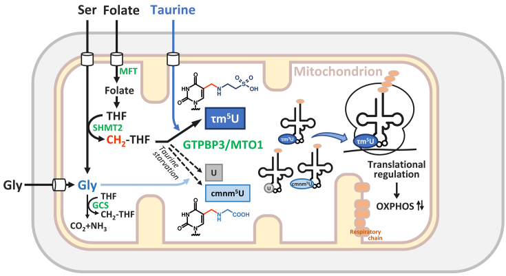
Modified uridine containing taurine, 5-taurinomethyluridine (τm5U), is found at the anticodon first position of mitochondrial (mt-)transfer RNAs (tRNAs). Previously, we reported that τm5U is absent in mt-tRNAs with pathogenic mutations associated with mitochondrial diseases. However, biogenesis and physiological role of τm5U remained elusive. Here, we elucidated τm5U biogenesis by confirming that 5,10-methylene-tetrahydrofolate and taurine are metabolic substrates for τm5U formation catalyzed by MTO1 and GTPBP3. GTPBP3-knockout cells exhibited respiratory defects and reduced mitochondrial translation. Very little τm5U34 was detected in patient's cells with the GTPBP3 mutation, demonstrating that lack of τm5U results in pathological consequences. Taurine starvation resulted in downregulation of τm5U frequency in cultured cells and animal tissues (cat liver and flatfish). Strikingly, 5-carboxymethylaminomethyluridine (cmnm5U), in which the taurine moiety of τm5U is replaced with glycine, was detected in mt-tRNAs from taurine-depleted cells. These results indicate that tRNA modifications are dynamically regulated via sensing of intracellular metabolites under physiological condition.


Figures

Figure 1.
Figure 1.
Metabolic labeling of mt-tRNAs to determine metabolic sources of τm5U. (A) Chemical structure of τm5(s2)U which is present at the wobble (first) position of anticodon of five mt-tRNAs. (B) Scheme for metabolic labeling experiments. HeLa cells were cultured with stable isotope (SI)-labeled or non-labeled amino acids. Total RNA was extracted from each culture, and individual mt-tRNAs were isolated and digested into nucleosides, followed by liquid chromatography–mass spectrometry (LC/MS) analysis to detect SI-labeled τm5(s2)U. (C) Mass spectra of τm5U in mt-tRNATrp isolated from HeLa cells cultured in normal medium (first from the left), [18O] taurine–supplemented medium (second) and [15N] taurine–supplemented medium (third). Labeled atoms are colored as indicated. Increased m/z values of τm5U due to the presence of SI-atoms are shown in parentheses. (D) Mass spectra of τm5U in mt-tRNAs for Trp and Leu(UUR) isolated from HeLa cells cultured in [15N, 13C3] Ser–supplemented medium (left) and [13C] Gly–supplemented medium (right). 13C atoms in their chemical structures are indicated by asterisks. (E) Collision-induced dissociation (CID) spectrum of [13C]-labeled τm5U nucleoside. The precursor ion (m/z 383.1) for CID is marked by an arrow. Product ions in the CID spectrum are assigned in the chemical structure of τm5U. (F) Mitochondrial 1C metabolism involved in τm5U biosynthesis (see Figure 7). Extracted mass chromatograms (XICs) of anticodon-containing RNase T1-digested fragments bearing U34 (upper panels) or τm5U34 (lower panels) from mt tRNALeu(UUR) isolated from Chinese hamster ovary (CHO) wild-type (WT) (left panels), Shmt2 mutant (center panels) and Mft mutant (right panels) cells. Sequence of the detected fragment, with its m/z value and charge state, is indicated on the right.
Figure 2.
Figure 2.
Downregulation of τm5(s2)U in culture cells and animal tissues upon taurine starvation. (A) XICs of anticodon-containing RNase T1-digested fragments with different wobble modifications of mt-tRNALys isolated from HeLa cells cultured in normal (left panels), taurine-depleted (center panels) and taurine-restoring (right panels) conditions. Sequence of the detected fragment, with its m/z value and charge state, is indicated on the right. Relative τm5(s2)U frequencies indicated in the bottom chromatograms are averaged values with standard deviations, calculated from the peak height ratio of the multiply charged negative ions (−3 to −7) for RNA fragments with different modifications. s2U, 2-thiouridine; t6A, N6-threonylcarbamoyladenosine. (B) Averaged mass spectra of the anticodon-containing fragments with different wobble modifications of mt-tRNALys isolated from HeLa cells cultured in normal (left panel) and taurine-depleted (right panel) conditions. (C) CID spectrum of the anticodon-containing RNase T1-digested fragment bearing cmnm5s2U of mt-tRNAGln isolated from HeLa cells cultured under taurine-depleted conditions. The c- and y-series of the product ions are assigned on the sequence. (D) XICs of anticodon-containing RNase T1-digested fragments with different wobble modifications of mt-tRNALys, isolated from livers of cats fed a normal diet (left panels) or a taurine-depleted diet (right panels). Sequence of the detected fragment, with its m/z value and charge state, is indicated on the right. Relative τm5(s2)U frequencies indicated in the bottom chromatograms are averaged values with standard deviations, calculated from the peak height ratio of the multiply charged negative ions (−3 to −7) for RNA fragments with different modifications. (E) XICs of anticodon-containing RNase T1-digested fragments with different wobble modifications of mt-tRNALys, isolated from muscles of flatfish fed a normal diet (left panels) or a taurine-depleted diet (right panels). Sequence of the detected fragment, with its m/z value and charge state, is indicated on the right. Relative τm5(s2)U frequencies indicated in the bottom chromatograms are averaged values with standard deviations, calculated from the peak height ratio of the multiply charged negative ions (−2 to −4) for RNA fragments with different modifications.
Figure 3.
Figure 3.
GTPBP3 and MTO1 are essential for τm5U biogenesis. (A) A diagram of the human GTPBP3 gene and the sites of mutations introduced by the CRISPR–Cas9 system. Shaded boxes indicate coding regions, open boxes indicate untranslated regions of exons and lines indicate introns. Heterozygous mutations found in the patient with mitochondrial disease are indicated above the diagram (34,35). The sgRNA sequence is underlined; the PAM sequence is in red. Reading frame and translated amino acids are shown below the sequence. (B) Genomic PCR products covering the sgRNA target site from WT HEK293T and GTPBP3 KO cells. One allele of the KO cell has a 318 bp insertion, and the other has a 74 bp deletion. (C) Western blotting of GTPBP3 and actin proteins in WT and KO cells. (D) Steady-state levels of six mt-tRNAs from WT and KO cells, evaluated by northern blotting. The bar graph shows relative amounts of mt-tRNALys from WT and KO cells, quantified in three independent replicates. Error bars indicate standard deviation. *p = 0.02, Student’s paired t-test. (E) XICs of anticodon-containing RNase T1-digested fragments with U34 (upper panels) and τm5U34 (lower panels) of mt tRNALeu(UUR), isolated from WT (left panels) and GTPBP3 KO cells (right panels). Sequence of the detected fragment, with its m/z value and charge state, is indicated on the right. (F) XICs of anticodon-containing RNase T1-digested fragments with U34 (upper panels) and τm5U34 (lower panels) of mt tRNALeu(UUR), isolated from WT (left panels) and Mto1 KO mES cells (right panels). Sequence of the detected fragment, with its m/z value and charge state, is indicated on the right.
Figure 4.
Figure 4.
Mitochondrial dysfunction in GTPBP3 KO cells. (A) Oxygen consumption rate in HEK293T WT (square) and GTPBP3 KO (circle) cells, measured using a flux analyzer. Each plot represents the mean value of three independent samples. Error bars indicate standard deviations. (B) Relative activities of respiratory chain complexes I–IV and citrate synthase of WT (black bar) and GTPBP3 KO (gray bar) cells. Error bars indicate standard deviation. *p < 0.05, **p< 0.01, Student’s t-test. (C) Western blotting of subunit proteins (indicated on the right) of respiratory chain complexes in isolated mitochondria from WT and GTPBP3 KO cells. The CBB-stained gel images are shown in Supplementary Figure S8A. (D) Pulse-labeling of mitochondrial protein synthesis. WT and GTPBP3 KO cells were labeled with [35S] Met and [35S] Cys after cytoplasmic protein synthesis was halted by emetine. Assignment of mitochondrial proteins is indicated on the left. The CBB-stained gel images are shown in Supplementary Figure S8B.
Figure 5.
Figure 5.
Hypomodification of the taurine modification in a patient with pathogenic mutations in GTPBP3. XICs of anticodon-containing RNase T1-digested fragments with different modifications of mt-tRNAs for Leu(UUR) (A), Trp (B), Gln (C), Glu (D) and Lys (E), isolated from neonatal human dermal fibroblast (NHDF) (left panels) and patient fibroblasts (right panels). Sequence of the detected fragment, with its m/z value and charge state, is indicated on the right. Relative frequencies of τm5(s2)U in the patient sample are indicated in the bottom chromatograms. n.d., not detected. ms2i6A, 2-methylthio-N6-isopentenyladenosine; i6A, N6-isopentenyladenosine.
Figure 6.
Figure 6.
In vitro reconstitution of τm5U34. (A) In vitro reconstitution of cmnm5U34 and τm5U34 on E. coli tRNAGly(UCC) in the presence of Gly (left panels) or taurine (right panels) along with other substrates and co-factors. XICs of anticodon-containing RNase T1-digested fragments with U34 (top panels), cmnm5U34 (middle panels) and τm5U34 (bottom panels) of E. coli tRNAGly(UCC) after the reaction are shown. Sequence of the detected fragment, with its m/z value and charge state, is indicated on the right. (B) Mass spectrum of the anticodon-containing RNase T1-digested fragment of E. coli tRNAGly(UCC) containing τm5U34, reconstituted in vitro. The exact mass of the mono-isotopic ion is indicated. (C) CID spectrum of the above fragment with τm5U34. Assigned c- and y-product ions are indicated. (D) Co-immunoprecipitation of GTPBP3 and MTO1-FLAG complex. Both proteins were transiently co-expressed in HEK293T cells, pulled down with anti-FLAG antibody–agarose and eluted with FLAG peptide. Whole lysate and eluate were resolved by SDS-PAGE and stained with a fluorescent dye. MTS stands for mitochondrial targeting sequence. (E) In vitro reconstitution of τm5U on mt-tRNALeu(UUR) without enzymes (left panels), without GTP and taurine (middle panels), or with all components including substrates and co-factors (right panels). XICs of anticodon-containing RNase T1-digested fragments with U34 (top panels) and τm5U34 (bottom panels) of mt-tRNALeu(UUR) after the reaction. Sequence of the detected fragment, with its m/z value and charge state, is indicated on the right. (F) Scheme of τm5U and cmnm5U biosynthesis. τm5U is synthesized by GTPBP3/MTO1 complex using 5,10-CH2-THF and taurine as substrates while glycine is used in place of taurine for cmnm5U by MnmE–MnmG complex. Co-factors including GTP, FAD and potassium ion are essential to cmnm5U formation, suggesting that they are also required for τm5U.
Figure 7.
Figure 7.
Metabolic regulation and chemical switching of tRNA modification in mitochondria. Schematic depiction of 1C metabolism, τm5U biogenesis and potential translational regulation in mitochondria. τm5U is biosynthesized from CH2-THF and taurine catalyzed by GTPBP3–MTO1 complex. τm5U in mt-tRNAs is required for efficient mitochondrial translation to synthesize the respiratory chain complexes essential for OXPHOS on the mitochondrial inner membrane. Taurine starvation decreases τm5U frequency and increases unmodified U. In this condition, cmnm5U, in which the taurine moiety of τm5U is replaced with glycine, is also introduced in mt-tRNAs. The tRNA modification dynamically regulated in response to metabolic status implies translational regulation in a codon-specific manner under physiological conditions. Abbreviations are as follows: THF, tetrahydrofolate; SHMT2, serine hydroxymethyltransferase 2 (mitochondrial SHMT); MFT, mitochondrial folate transporter; GCS, glycine cleavage system.

References

    1. Machnicka M.A., Milanowska K., Osman Oglou O., Purta E., Kurkowska M., Olchowik A., Januszewski W., Kalinowski S., Dunin-Horkawicz S., Rother K.M. et al. MODOMICS: a database of RNA modification pathways–2013 update. Nucleic Acids Res. 2013; 41:D262–D267. - PMC - PubMed
    1. Chen K., Zhao B.S., He C.. Nucleic acid modifications in regulation of gene expression. Cell Chem. Biol. 2016; 23:74–85. - PMC - PubMed
    1. Gilbert W.V., Bell T.A., Schaening C.. Messenger RNA modifications: form, distribution and function. Science. 2016; 352:1408–1412. - PMC - PubMed
    1. Frye M., Jaffrey S.R., Pan T., Rechavi G., Suzuki T.. RNA modifications: what have we learned and where are we headed. Nat. Rev. Genetics. 2016; 17:365–372. - PubMed
    1. Duechler M., Leszczynska G., Sochacka E., Nawrot B.. Nucleoside modifications in the regulation of gene expression: focus on tRNA. Cell Mol. Life Sci. 2016; 73:3075–3095. - PMC - PubMed

Publication types