

An ethmoid mucocele causing diplopia: A case report
- PMID: 29390410
- PMCID: PMC5815822
- DOI: 10.1097/MD.0000000000009353
An ethmoid mucocele causing diplopia: A case report
Abstract
Rationale: Mucocele is a disease lined primarily by epithelium, and occurs mainly when the sinus ostium is obstructed.
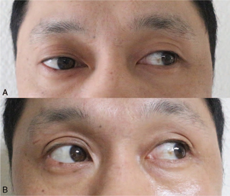
Patient concerns: We report a case of a 37-year-old man who presented with painless proptosis of the right eye and diplopia.
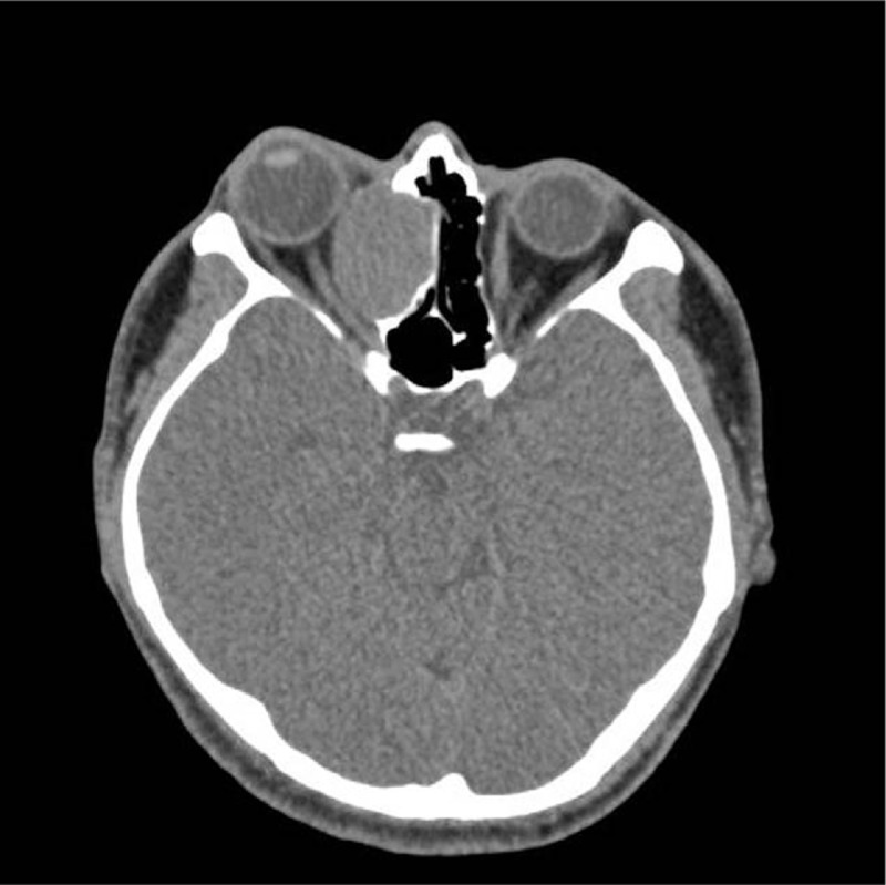
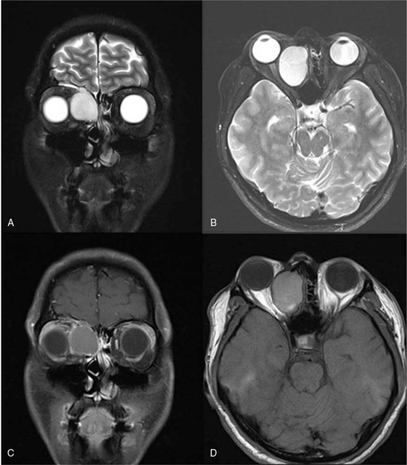
Diagnoses: The preoperative finding was mucocele of the ethmoid sinus.
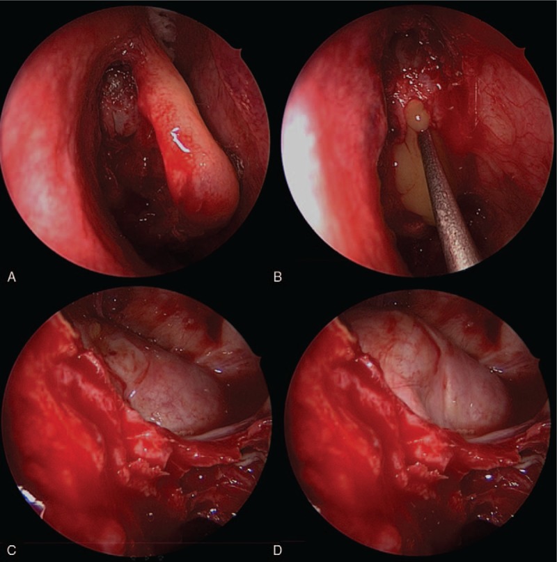
Interventions: We performed endoscopic sinus surgery, which included uncapping of the anterior and superior wall of the mucocele.
Outcomes: The mucocele was treated safely and effectively without touching the medial orbital wall.
Lessons: Clinicians should note that minimally invasive surgery to remove ethmoid mucoceles is relatively straightforward and can prevent the various complications associated with these lesions.
Copyright © 2017 The Authors. Published by Wolters Kluwer Health, Inc. All rights reserved.
Conflict of interest statement
The authors report no conflict of interest.
Figures
References
-
- Verillaud B, Le Clerc N, Blancal JP, et al. Mucocele formation after surgical treatment of inverted papilloma of the frontal sinus drainage pathway. Am J Rhinol Allergy 2016;30:181–4. - PubMed
-
- Tseng CC, Ho CY, Kao SC. Ophthalmic manifestations of paranasal sinus mucoceles. J Chin Med Assoc 2005;68:260–4. - PubMed
-
- Morita S, Mizoguchi K, Iizuka K. Paranasal sinus mucoceles with visual disturbance. Auris Nasus Larynx 2010;37:708–12. - PubMed
Publication types
MeSH terms
LinkOut - more resources
Full Text Sources
Other Literature Sources
