

Circumferential Esophageal Replacement by a Tissue-engineered Substitute Using Mesenchymal Stem Cells: An Experimental Study in Mini Pigs
- PMID: 29390879
- PMCID: PMC5802636
- DOI: 10.1177/0963689717741498
Circumferential Esophageal Replacement by a Tissue-engineered Substitute Using Mesenchymal Stem Cells: An Experimental Study in Mini Pigs
Abstract
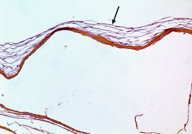
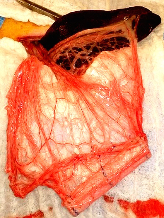
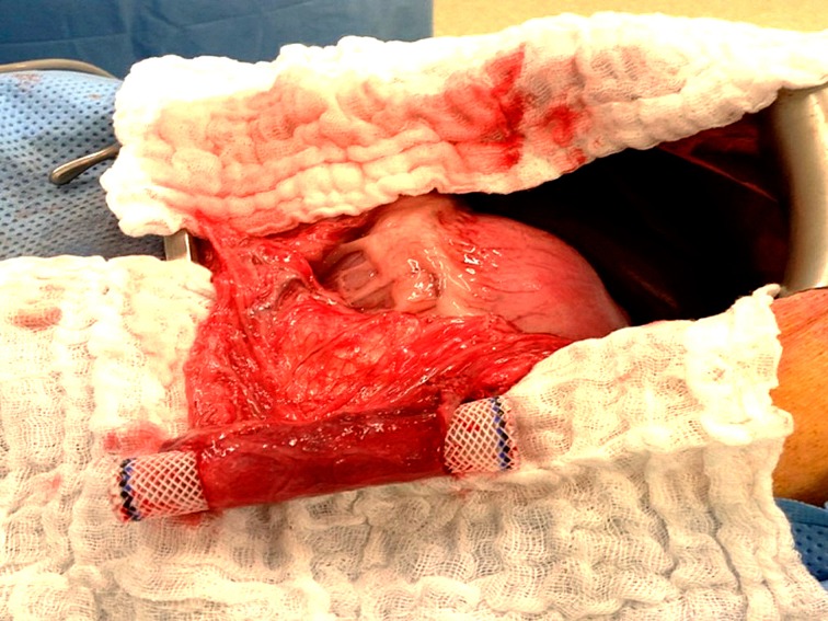
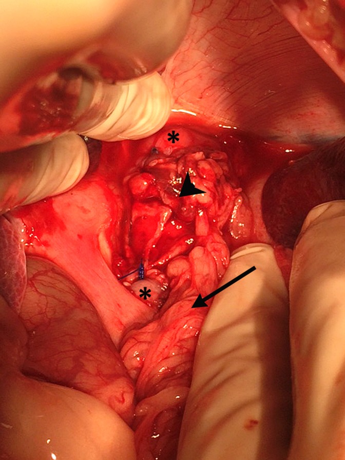
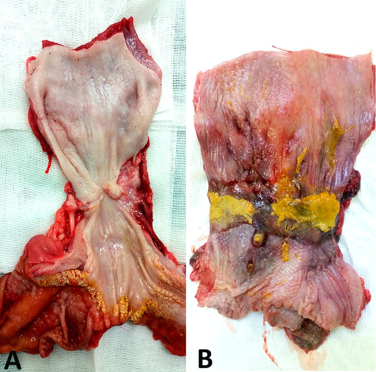
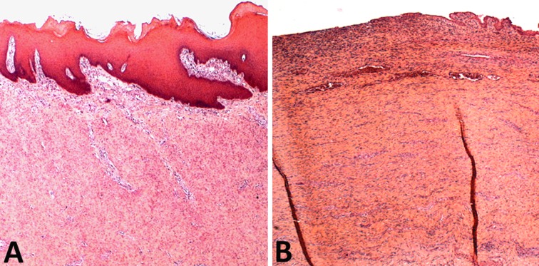
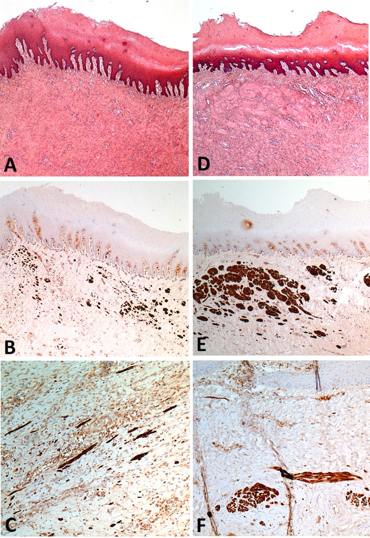
Tissue engineering appears promising as an alternative technique for esophageal replacement. Mesenchymal stem cells (MSCs) could be of interest for esophageal regeneration. Evaluation of the ability of an acellular matrix seeded with autologous MSCs to promote tissue remodeling toward an esophageal phenotype after circumferential replacement of the esophagus in a mini pig model. A 3 cm long circumferential replacement of the abdominal esophagus was performed with an MSC-seeded matrix (MSC group, n = 10) versus a matrix alone (control group, n = 10), which has previously been matured into the great omentum. The graft area was covered with an esophageal removable stent. A comparative histological analysis of the graft area after animals were euthanized sequentially is the primary outcome of the study. Histological findings after maturation, overall animal survival, and postoperative morbidity were also compared between groups. At postoperative day 45 (POD 45), a mature squamous epithelium covering the entire surface of the graft area was observed in all the MSC group specimens but in none of the control group before POD 95. Starting at POD 45, desmin positive cells were seen in the graft area in the MSC group but never in the control group. There were no differences between groups in the incidence of surgical complications and postoperative death. In this model, MSCs accelerate the mature re-epitheliazation and early initiation of muscle cell colonization. Further studies will focus on the use of cell tracking tools in order to analyze the becoming of these cells and the mechanisms involved in this tissue regeneration.
Keywords: acellular matrix; esophageal stenting; esophagus; large animal model; mesenchymal stem cells; tissue engineering; translational research.
Conflict of interest statement
Figures
References
-
- Poghosyan T, Gaujoux S, Chirica M, Munoz-Bongrand N, Sarfati E, Cattan P. Functional disorders and quality of life after esophagectomy and gastric tube reconstruction for cancer. J Visc Surg. 2011;148(5):327–335. - PubMed
-
- Chirica M, Veyrie N, Munoz-Bongrand N, Zohar S, Halimi B, Celerier M, Cattan P, Sarfati E. Late morbidity after colon interposition for corrosive esophageal injury: risk factors, management, and outcome. A 20-years experience. Ann Surg. 2010;252(2):271–280. - PubMed
-
- Poghosyan T, Catry J, Luong-Nguyen M, Bruneval P, Domet T, Arakelian L, Sfeir R, Michaud L, Vanneaux V, Gottrand F, et al. Esophageal tissue engineering: current status and perspectives. J Visc Surg. 2016;153(1):21–29. - PubMed
-
- Saxena AK. Esophagus tissue engineering: designing and crafting the components for the “hybrid construct” approach. Eur J Pediatr Surg. 2014;24(3):246–262. - PubMed
-
- Poghosyan T, Sfeir R, Michaud L, Bruneval P, Domet T, Vanneaux V, Luong-Nguyen M, Gaujoux S, Gottrand F, Larghero J, et al. Circumferential esophageal replacement using a tube-shaped tissue-engineered substitute: An experimental study in minipigs. Surgery. 2015;158(1):266–277. - PubMed
Publication types
MeSH terms
LinkOut - more resources
Full Text Sources
Other Literature Sources
