Clinical Trial
. 2018 Jun;138(6):1301-1310.
doi: 10.1016/j.jid.2018.01.006. Epub 2018 Jan 31.

Mycophenolate Mofetil Treatment of Systemic Sclerosis Reduces Myeloid Cell Numbers and Attenuates the Inflammatory Gene Signature in Skin

Affiliations
Clinical Trial

Mycophenolate Mofetil Treatment of Systemic Sclerosis Reduces Myeloid Cell Numbers and Attenuates the Inflammatory Gene Signature in Skin

Monique Hinchcliff et al. J Invest Dermatol. 2018 Jun.

Abstract

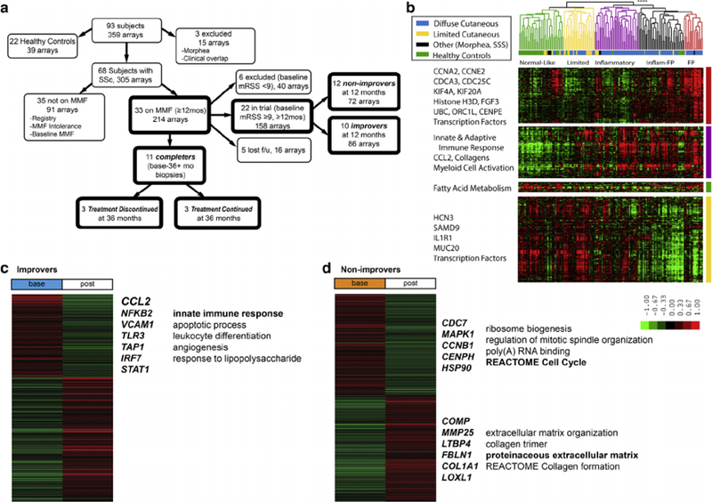
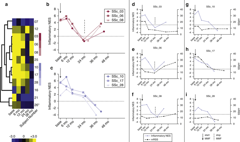
Fewer than half of patients with systemic sclerosis demonstrate modified Rodnan skin score improvement during mycophenolate mofetil (MMF) treatment. To understand the molecular basis for this observation, we extended our prior studies and characterized molecular and cellular changes in skin biopsies from subjects with systemic sclerosis treated with MMF. Eleven subjects completed ≥24 months of MMF therapy. Two distinct skin gene expression trajectories were observed across six of these subjects. Three of the six subjects showed attenuation of the inflammatory signature by 24 months, paralleling reductions in CCL2 mRNA expression in skin and reduced numbers of macrophages and myeloid dendritic cells in skin biopsies. MMF cessation at 24 months resulted in an increased inflammatory score, increased CCL2 mRNA and protein levels, modified Rodnan skin score rebound, and increased numbers of skin myeloid cells in these subjects. In contrast, three other subjects remained on MMF >24 months and showed a persistent decrease in inflammatory score, decreasing or stable modified Rodnan skin score, CCL2 mRNA reductions, sera CCL2 protein levels trending downward, reduction in monocyte migration, and no increase in skin myeloid cell numbers. These data summarize molecular changes during MMF therapy that suggest reduction of innate immune cell numbers, possibly by attenuating expression of chemokines, including CCL2.


Conflict of interest statement

CONFLICT OF INTEREST

Figures

Figure 1.
Figure 1.. Overview of study design and intrinsic gene expression analysis.
(a) Flowchart of subjects through clinical study, (b) 2,500 probes were selected with a false discovery rate of 0.14% from 163 baseline arrays by intrinsic gene analysis by subject, This hierarchical clustering dendrogram shows the normal-like (green), limited (yellow), inflammatory (purple), and fibroproliferative (red) groups. Significance of clustering was determined by SigClust (P ≤ 0.0001). (c) 651 genes in improvers and (d) 1,067 genes in non-improvers were differentially expressed between baseline and 12-month (post) biopsies (false discovery rate <5%; arm and flank biopsies used). The gene centroid (average of all arrays in a class) is displayed. Selected genes and pathways are displayed to the right of the centroid. Bolded gene symbols are annotated to the pathway/Gene Ontology term in bold.
Figure 2.
Figure 2.. The inflammatory signature is attenuated by MMF but rebounds in subjects that cease MMF treatment at 24 months.
Single-sample Gene Set Enrichment Analysis normalized enrichment scores (NES) were used to summarize changes in inflammatory gene expression during MMF treatment. (a) The inflammatory NES of completers were hierarchically clustered and displayed as a heatmap and (b, c) as line plots for select subjects. (d – f) Three subjects lose their inflammatory signature during treatment and rebound at 36 months after treatment cessation. (g – i) Three subjects who continue treatment do not show an increase in inflammatory signature. An arrow indicates when treatment began. A dashed line indicates when treatment stopped. The purple line indicates NES. The black dashed line indicates mRSS. *Subject 26 was never on MMF, and serves as a control. MMF, mycophenolate mofetil; mRSS, modified Rodnan skin score; SSc, systemic sclerosis.
Figure 3.
Figure 3.. Changes in CCL2 expression in skin, CCL2 concentration in sera, and monocyte migration of SSc patients treated with MMF.
CCL2 mRNA levels were quantified using quantitative real-time PCR and normalized to beta-actin mRNA in (a). Treatment Discontinued and (b) Treatment Continued groups. (c) ELISA was used to measure circulating CCL2 of SSc patients at baseline (n = 3), 3 months of MMF (n = 2), and >16 months of MMF (n = 4) and healthy controls (n = 4, females, >30 years) in pg/ml. (d) Relative monocyte migration toward sera from SSc patients and sera or plasma from healthy controls (n = 5, females, >30 years) was measured. Relative fluorescence (y-axis) is the ratio of sample fluorescence and internal control fluorescence (media without additional CCL2). Unpaired t test used for all statistical analysis, mean with standard deviation (SD) shown. MMF, mycophenolate mofetil; SSc, systemic sclerosis.
Figure 4.
Figure 4.. Myeloid dendritic cells, T-lymphocytes, and macrophages contribute significantly to the inflammatory gene expression signature.
(a) Six subjects highlighted in Figure 2 show a high correlation between their individual cell type-specific NES scores and their inflammatory NES scores across the time course for dendritic cells (DCs; r = 0.83), macrophages (r = 0.60), and T-lymphocytes (r = 0.77). (b, c) The cell type NES scores are plotted against time (base, 6 months, 12 months, 24 months, and 36 months) and are split between subjects who discontinue MMF at 24 months (b, Treatment Discontinued) and subjects who continue MMF treatment (c, Treatment Continued). Treatment Discontinued are subjects SSc_03, 06, and 08 and Treatment Continued are subjects SSc_10, 17, and 28. Arrow indicates when a subject started MMF, and a dashed line indicates when a subject discontinued MMF.MMF, mycophenolate mofetil; NES, normalized enrichment scores; SSc, systemic sclerosis.
Figure 5.
Figure 5.. Immunohistochemistry shows reductions in DCs and macrophages during MMF treatment.
(a, b) Immunohistochemistry staining targeting CD163 (macrophage marker) of arm biopsy slides. CD163 counts were plotted over time for subjects 03, 06, 08, 10, 17, and 28. (c, d) Immunohistochemistry staining for CD163 in baseline (c) and 36 months (d) biopsies from SSc_1 7 (Treatment Continued). (e, f) Immunohistochemistry staining targeting CD1c (mDC marker) of arm biopsy slides. CD1c counts were plotted overtime for the same subjects. (g–i) Immunohistochemistry staining for CD1c in baseline (g), 24-month (h), and 36-month (i) biopsies from SSc_08 (MMF ceased at 24 months, Treatment Discontinued). Data not available for all subjects at all time points; missing data denoted by “x.” An arrow indicates when treatment started. A dashed line indicates when treatment stopped. DC, dendritic cell; mDC, myeloid dendritic cell; MMF, mycophenolate mofetil.

References

    1. Assassi S, Swindell WR, Wu M, Tan FD, Khanna D, Furst DE, et al. Dissecting the heterogeneity of skin gene expression patterns in systemic sclerosis. Arthritis Rheumatol 2015;67:3016–26. - PMC - PubMed
    1. Assassi S, Wu M, Tan FK, Chang J, Graham TA, Furst DE, et al. Skin gene expression correlates of severity of interstitial lung disease in systemic sclerosis. Arthritis Rheum 2013;65:2917–27. - PMC - PubMed
    1. Bolen CR, Uduman M, Kleinstein SH. Cell subset prediction for blood genomic studies. BMC Bioinform 2011;12:258. - PMC - PubMed
    1. Chia JJ, Lu TT. Update on macrophages and innate immunity in scleroderma. Curr Opin Rheumatol 2015;27:530–6. - PMC - PubMed
    1. Christmann RB, Hayes E, Pendergrass S, Padilla C, Farina G, Affandi AJ, et al. Interferon and alternative activation of monocyte/macrophages in systemic sclerosis-associated pulmonary arterial hypertension. Arthritis Rheum 2011;63:1718–28. - PMC - PubMed

Publication types

MeSH terms