. 2018 Feb 1;8(1):2117.
doi: 10.1038/s41598-018-20331-2.

Large-scale intact glycopeptide identification by Mascot database search

Affiliations

Large-scale intact glycopeptide identification by Mascot database search

Ravi Chand Bollineni et al. Sci Rep. .

Erratum in

Abstract

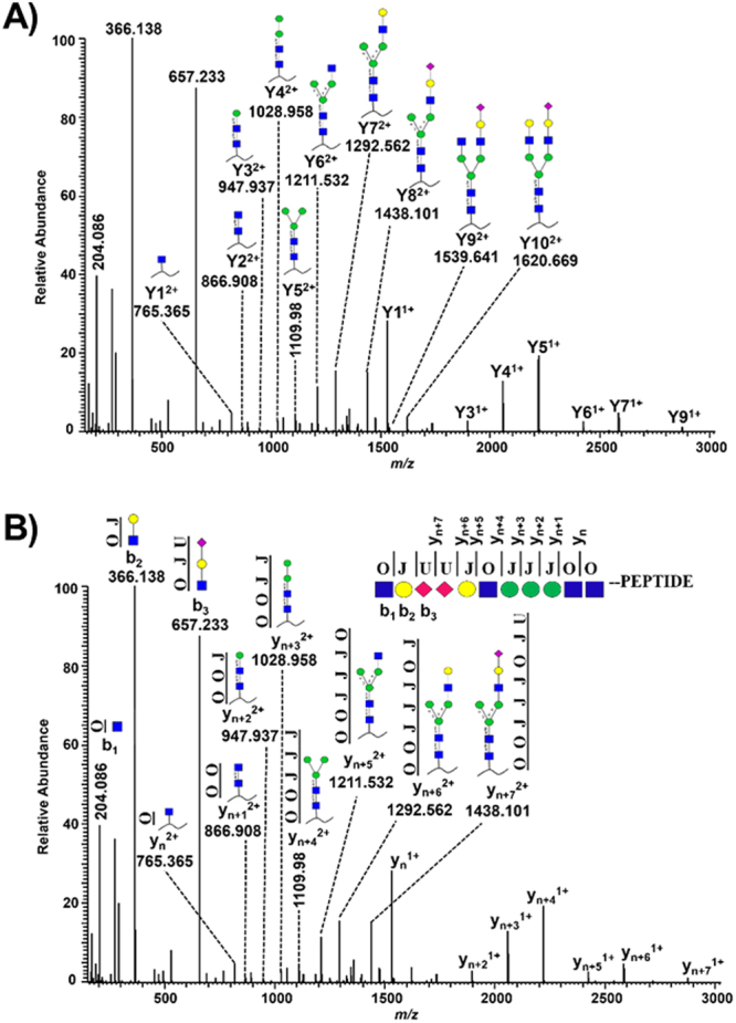
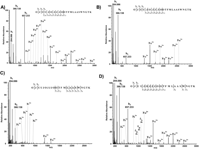
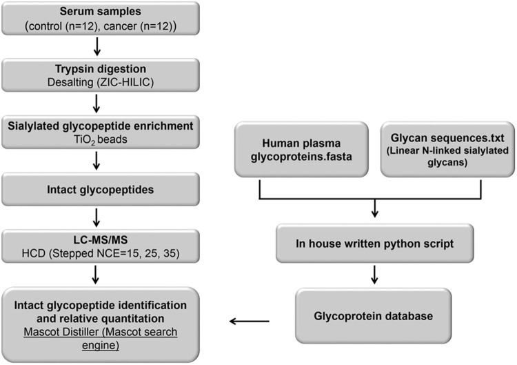
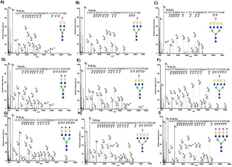
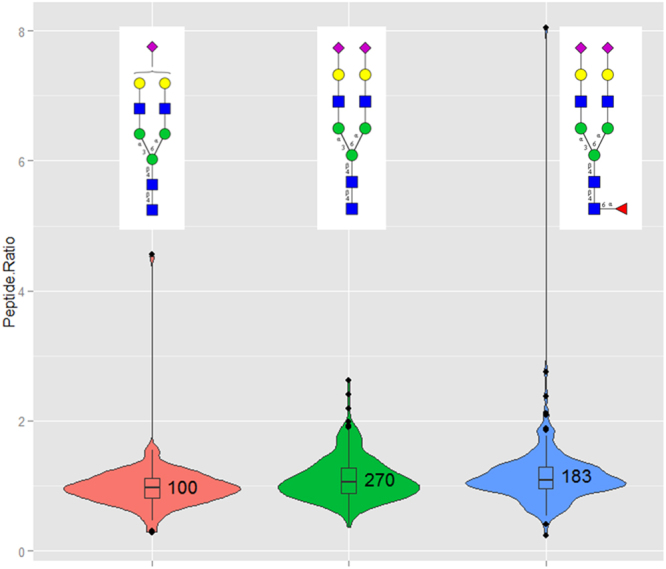
Workflows capable of determining glycopeptides in large-scale are missing in the field of glycoproteomics. We present an approach for automated annotation of intact glycopeptide mass spectra. The steps in adopting the Mascot search engine for intact glycopeptide analysis included: (i) assigning one letter codes for monosaccharides, (ii) linearizing glycan sequences and (iii) preparing custom glycoprotein databases. Automated annotation of both N- and O-linked glycopeptides was proven using standard glycoproteins. In a large-scale study, a total of 257 glycoproteins containing 970 unique glycosylation sites and 3447 non-redundant N-linked glycopeptide variants were identified in 24 serum samples. Thus, a single tool was developed that collectively allows the (i) elucidation of N- and O-linked glycopeptide spectra, (ii) matching glycopeptides to known protein sequences, and (iii) high-throughput, batch-wise analysis of large-scale glycoproteomics data sets.


Conflict of interest statement

The authors declare no competing interests.

Figures

Figure 1
Figure 1
HCD-MS2 spectrum of a di-sialylated bi-antennary glycopeptide (m/z 1177.81373+) derived from bovine alpha-1-acid glycoprotein 1. The glycopeptide was fragmented at an NCE value of 15. (A) Yn represent the peptide bound glycosidic cleavage ions and the insert shows the corresponding peptide bound glycan structures. (B) Linearizing glycan structures with the corresponding three letters O (GlcNAc, GalNAc), J (Galactose, Mannose) and U (Neu5Ac) are depicted. A linearized di-sialylated bi-antennary glycan structure attached to the N-terminus of a peptide and annotated y and b type cleavage ions corresponds to the MS2 spectrum.
Figure 2
Figure 2
Mascot annotated MS2 spectra of a di-sialylated bi-antennary N-glycopeptide (m/z 1177.81373+) fragmented at different NCE values. NCE values of (A) 15, (B) 25, (C) 35 and (D) the composite MS2 spectrum of the same precursor fragmented using the stepped NCE values of 15, 25 and 35 are displayed. Standard bovine alpha-1-acid glycoprotein 1 was digested with trypsin, the glycopeptides were analyzed by LC-MS and the data were searched against the custom glycoprotein database using the Mascot search engine.
Figure 3
Figure 3
Mascot annotated O-glycopeptide MS2 spectra of fetuin using stepped NCE values. Bovine fetuin was digested with trypsin, analyzed by LC-MS using the stepped NCE function (15, 25 and 35) and searched against the custom O-glycoprotein database. Mascot annotated mono- (A,C) and di-sialylated (B,D) core-1 O-linked glycopeptide spectra from two different peptide sequences.
Figure 4
Figure 4
Workflow for N-glycoproteome analyses of serum samples. Briefly, the serum samples from control and patients diagnosed with prostate cancer were digested with trypsin and desalted with ZIC-HILIC SPE. Next, the N-linked sialylated glycopeptides were enriched with TiO2 beads, followed by LC-MS analysis using a Q Exactive mass spectrometer applying stepped NCE for HCD fragmentation. The intact glycopeptide mass spectra were submitted to the Mascot search engine for identification and relative quantification with Mascot Distiller. The data was searched against a custom glycoprotein database prepared from 21 linear N-linked sialylated glycans and proteins (444) known to be glycosylated in serum (PeptideAtlas N-Glyco build 2010).
Figure 5
Figure 5
Annotation of nine different glycan structures with varied degree of complexity and sialylation by Mascot on a single glycosylation site (Asn 93) of alpha-1-acid glycoprotein 1 in serum. Shown here are the representative HCD MS2 spectra annotated by Mascot. The nine different glycopeptide variants included the mono-sialylated bi- (A), tri- (B), tetra-antennary (C), and the di-sialylated bi- (D), tri- (E), tetra-antennary (F). Tri- (G,H) and tetra-sialylated (I) glycan structures on the same glycosylation site were also annotated by Mascot.
Figure 6
Figure 6
Violin plots representing the glycopeptide ratios (aggressive vs. indolent prostate cancer) of the three most frequent glycopeptide variants identified in the current study. Glycopeptides identified and quantified in 24 serum samples were segmented based on the glycan structures irrespective of the protein origin. The three most frequent glycopeptide variants were the mono-sialylated bi-antennary, di-sialylated bi-antennary without and with one fucose residue. Mascot Distiller was used to calculate the XIC values and the corresponding ratios between aggressive (12) and indolent (12) samples. Glycopeptide precursors contributing to a minimum 50% of the XIC peak area and passing the correlation threshold of 0.8 were only considered.

References

    1. Marino K, Bones J, Kattla JJ, Rudd PM. A systematic approach to protein glycosylation analysis: a path through the maze. Nat Chem Biol. 2010;6:713–723. doi: 10.1038/nchembio.437. - DOI - PubMed
    1. Apweiler R, Hermjakob H, Sharon N. On the frequency of protein glycosylation, as deduced from analysis of the SWISS-PROT database. Biochim Biophys Acta. 1999;1473:4–8. doi: 10.1016/S0304-4165(99)00165-8. - DOI - PubMed
    1. Ohtsubo K, Marth JD. Glycosylation in cellular mechanisms of health and disease. Cell. 2006;126:855–867. doi: 10.1016/j.cell.2006.08.019. - DOI - PubMed
    1. Helenius A, Aebi M. Intracellular functions of N-linked glycans. Science. 2001;291:2364–2369. doi: 10.1126/science.291.5512.2364. - DOI - PubMed
    1. Xu C, Ng DT. Glycosylation-directed quality control of protein folding. Nat Rev Mol Cell Biol. 2015;16:742–752. doi: 10.1038/nrm4073. - DOI - PubMed

Publication types