. 2018 Feb 1;8(1):2082.
doi: 10.1038/s41598-018-19628-z.

Uptake, distribution, clearance, and toxicity of iron oxide nanoparticles with different sizes and coatings

Affiliations

Uptake, distribution, clearance, and toxicity of iron oxide nanoparticles with different sizes and coatings

Qiyi Feng et al. Sci Rep. .

Abstract

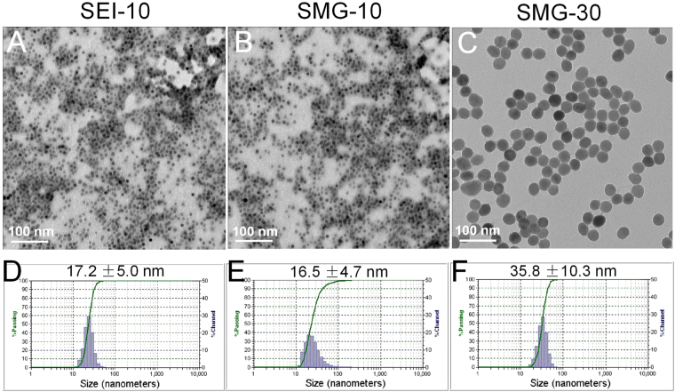
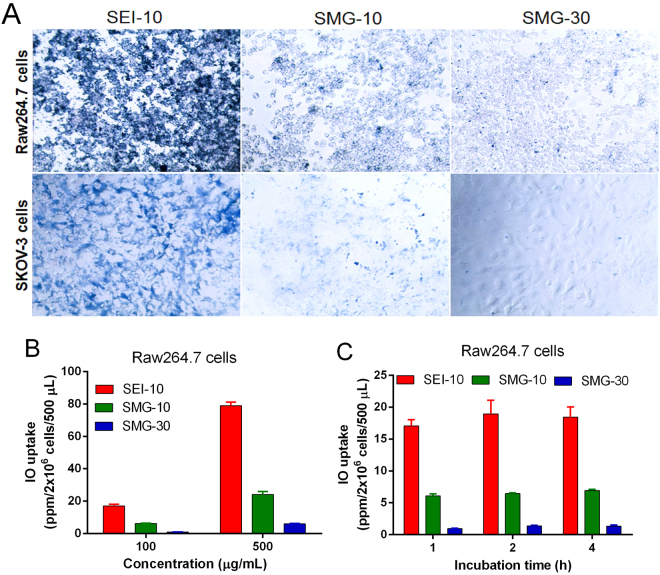
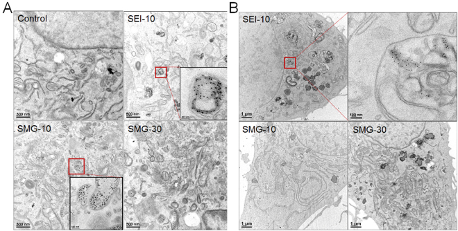
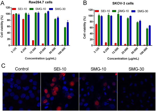
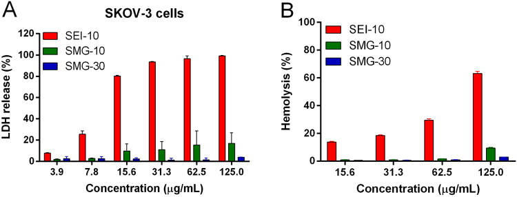
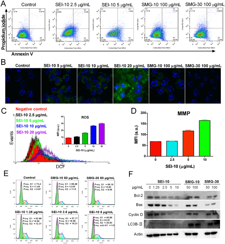
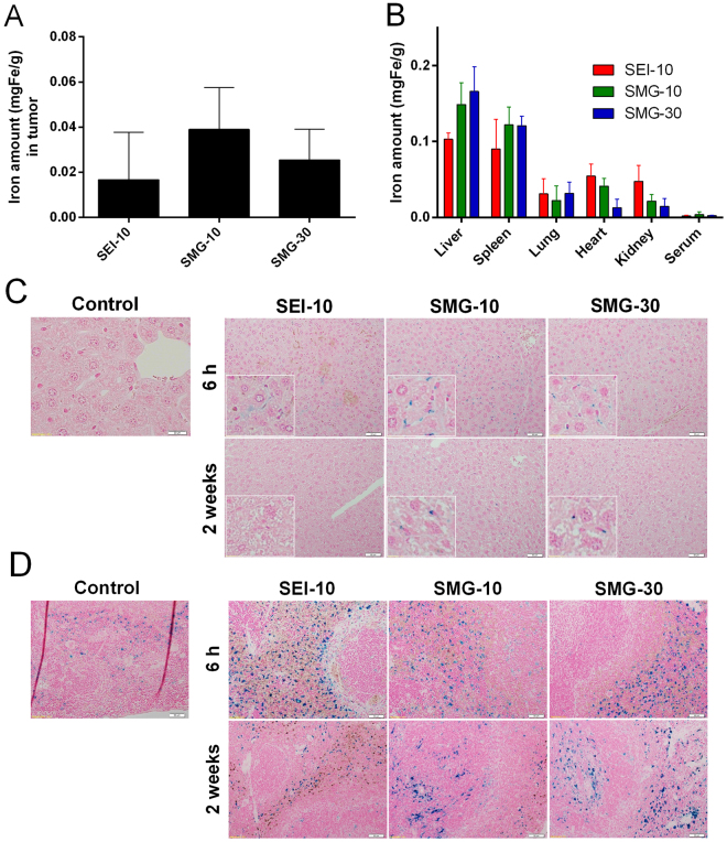
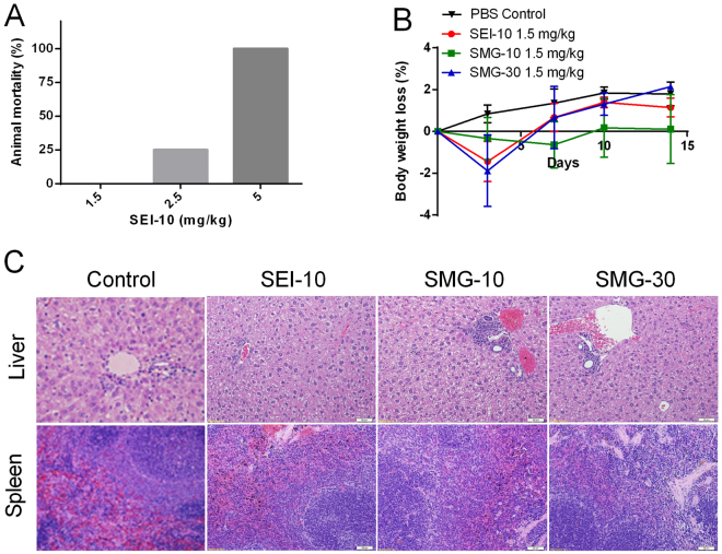
Iron oxide nanoparticles (IONPs) have been increasingly used in biomedical applications, but the comprehensive understanding of their interactions with biological systems is relatively limited. In this study, we systematically investigated the in vitro cell uptake, cytotoxicity, in vivo distribution, clearance and toxicity of commercially available and well-characterized IONPs with different sizes and coatings. Polyethylenimine (PEI)-coated IONPs exhibited significantly higher uptake than PEGylated ones in both macrophages and cancer cells, and caused severe cytotoxicity through multiple mechanisms such as ROS production and apoptosis. 10 nm PEGylated IONPs showed higher cellular uptake than 30 nm ones, and were slightly cytotoxic only at high concentrations. Interestingly, PEGylated IONPs but not PEI-coated IONPs were able to induce autophagy, which may play a protective role against the cytotoxicity of IONPs. Biodistribution studies demonstrated that all the IONPs tended to distribute in the liver and spleen, and the biodegradation and clearance of PEGylated IONPs in these tissues were relatively slow (>2 weeks). Among them, 10 nm PEGylated IONPs achieved the highest tumor uptake. No obvious toxicity was found for PEGylated IONPs in BALB/c mice, whereas PEI-coated IONPs exhibited dose-dependent lethal toxicity. Therefore, it is crucial to consider the size and coating properties of IONPs in their applications.


Conflict of interest statement

The authors declare that they have no competing interests.

Figures

Figure 1
Figure 1
Representative TEM images (A,B,C) and dynamic light scanning (DLS) size distribution (D,E,F) of SEI-10, SMG-10, and SMG-30.
Figure 2
Figure 2
Cell uptake of different IONPs in RAW264.7 macrophage cells and SKOV-3 cells. (A) Prussian blue staining of iron in RAW264.7 cells and SKOV-3 cells after incubation with SEI-10, SMG-10, and SMG-30 at a concentration of 200 μg/mL for 4 h, respectively. Magnification, 20×. ICP-MS measurement of iron concentrations in RAW264.7 cells after incubation with IONPs at different concentrations for 1 h (B) or at a concentration of 100 μg/mL for different period of time (C).
Figure 3
Figure 3
TEM images of RAW264.7 macrophages (A) and SKOV-3 cells (B) after 2-h exposure of different IONPs at the concentration of 100 µg/mL.
Figure 4
Figure 4
The in vitro cytotoxicity of different IONPs. The cell viability of RAW264.7 macrophages (A) and SKOV-3 cells (B) after 48-h treatment of IONPs with different concentrations were measured by MTS assay. (C) Representative fluorescent microscopic images showing the cell death mode induced by various IONPs in SKOV-3 cells. The cells were treated with SEI-10 (5 µg/mL), SMG-10 (400 µg/mL), or SMG-30 (400 µg/mL) for 16 h, and then stained with Hoechst 33342 (blue) and PI (red).
Figure 5
Figure 5
The LDH toxicities and hemolytic activities of different IONPs. (A) LDH release in SKOV-3 cells incubated with different concentrations of IONPs for 4 h. (B) In vitro red blood cells (RBCs) lysis. IONPs were incubated with mouse erythrocyte suspension for 4 h at 37 °C. RBCs lysis was determined spectrophotometrically (Absorbance 540 nm) based on hemoglobin level. PBS was used as negative control, and Triton-100 was used as positive control. Data represent mean ± SEM (n = 3).
Figure 6
Figure 6
The potential mechanisms underlying the cell death induced by IONPs in SKOV-3 cells. (A) Apoptotic and necrotic cell death were analyzed by Annexin V and PI dual staining after IONPs treatment for 24 h in SKOV-3 cells. Intracellular reactive oxygen species (ROS) production in SKOV-3 cells treated with various IONPs for 18 h was detected using a H2DCFDA probe by confocal microscopy (B) and flow cytometry (C), respectively. (D) The measurement of membrane mitochondria potential (MMP) of SKOV-3 cells treated with different concentrations of SEI-10 for 18 h. (E) the effect of IONPs on the cell cycle in SKOV-3 cells after 24-h treatment. (F) The effects of IONPs on the expression of proteins involved in apoptosis (Bcl-2, Bax), cell cycle (Cyclin D), and autophagy (LC3B-II), determined by western blot analysis after 18-h treatment.
Figure 7
Figure 7
In vivo biodistribution and clearance of different IONPs (1.5 mgFe/kg) in SKOV-3 tumor bearing mice after intravenous injection. The distribution of IONPs in the tumors (A) and other indicated organs (B) were quantified by ICP-MS at 24 h post-injection. Histological sections showing distribution, degradation, and clearance of IONPs in the liver (C) and spleen (D). At 6 h and 2 weeks post-injection, the sections of liver and spleen tissues harvested from the mice were processed with Prussian blue and nuclear fast red staining. The control samples were tissues from animals that were not injected with IONPs. The insets in (C) indicate the magnified images showing the Kupffer cells.
Figure 8
Figure 8
In vivo toxicity of different IONPs in BALB/c mice. (A) The mortality of BALB/c mice (n = 4) receiving different doses of SEI-10. (B) The body weight change of mice injected with various IONPs at the dose of 1.5 mg/kg, compared with PBS control. (C) Representative histopathological images of liver and spleen. Livers and spleens were harvested from mice on 2 weeks after intravenous injection of various IONPs, and stained with hematoxylin and eosin. Slight mononuclear cell infiltration in the portal area of the liver could be identified in mice with SMG-10 and SMG-30. Splenic plasmacytosis was also noted in mice treated with SMG-30, which may represent a response to antigenic stimulation.

References

    1. Naqvi S, et al. Concentration-dependent toxicity of iron oxide nanoparticles mediated by increased oxidative stress. Int. J. Nanomedicine. 2010;5:983–989. doi: 10.2147/IJN.S13244. - DOI - PMC - PubMed
    1. Bobo D, Robinson KJ, Islam J, Thurecht KJ, Corrie SR. Nanoparticle-Based Medicines: A Review of FDA-Approved Materials and Clinical Trials to Date. Pharm. Res. 2016;33:2373–2387. doi: 10.1007/s11095-016-1958-5. - DOI - PubMed
    1. Bulte JW. In vivo MRI cell tracking: clinical studies. AJR.American J. Roentgenol. 2009;193:314–325. doi: 10.2214/AJR.09.3107. - DOI - PMC - PubMed
    1. Wang Y-XJ. Current status of superparamagnetic iron oxide contrast agents for liver magnetic resonance imaging. World J Gastroenterol. 2015;21:1–15. doi: 10.3748/wjg.v21.i1.1. - DOI - PMC - PubMed
    1. Gustafson HH, Holt-Casper D, Grainger DW, Ghandehari H, Grainger D. Nanoparticle Uptake: The Phagocyte Problem HHS Public Access. Nano Today. 2015;10:487–510. doi: 10.1016/j.nantod.2015.06.006. - DOI - PMC - PubMed

Publication types