

Kaempferol mitigates Endoplasmic Reticulum Stress Induced Cell Death by targeting caspase 3/7
- PMID: 29391535
- PMCID: PMC5794799
- DOI: 10.1038/s41598-018-20499-7
Kaempferol mitigates Endoplasmic Reticulum Stress Induced Cell Death by targeting caspase 3/7
Abstract
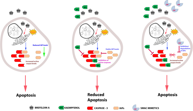
The Endoplasmic Reticulum (ER) plays a fundamental role in executing multiple cellular processes required for normal cellular function. Accumulation of misfolded/unfolded proteins in the ER triggers ER stress which contributes to progression of multiple diseases including neurodegenerative disorders. Recent reports have shown that ER stress inhibition could provide positive response against neuronal injury, ischemia and obesity in in vivo models. Our search towards finding an ER stress inhibitor has led to the functional discovery of kaempferol, a phytoestrogen possessing ER stress inhibitory activity in cultured mammalian cells. We have shown that kaempferol pre-incubation significantly inhibits the expression of GRP78 (a chaperone) and CHOP (ER stress associated pro-apoptotic transcription factor) under stressed condition. Also, our investigation in the inhibitory specificity of kaempferol has revealed that it inhibits cell death induced by diverse stimuli. Further study on exploring the molecular mechanism implied that kaempferol renders protection by targeting caspases. Both the in silico docking and in vitro assay using recombinant caspase-3 enzyme confirmed the binding of kaempferol to caspases, through an allosteric mode of competitive inhibition. Altogether, we have demonstrated the ability of kaempferol to alleviate ER stress in in vitro model.
Conflict of interest statement
The authors declare that they have no competing interests.
Figures


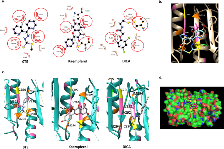
 ; Valine
  ; Methionine
  ) (d) Solid Surface representation of caspase-7 dimer with 0TE
  ; kaempferol
  ; and DICA
  ; docked poses superimposed at the dimerization site.
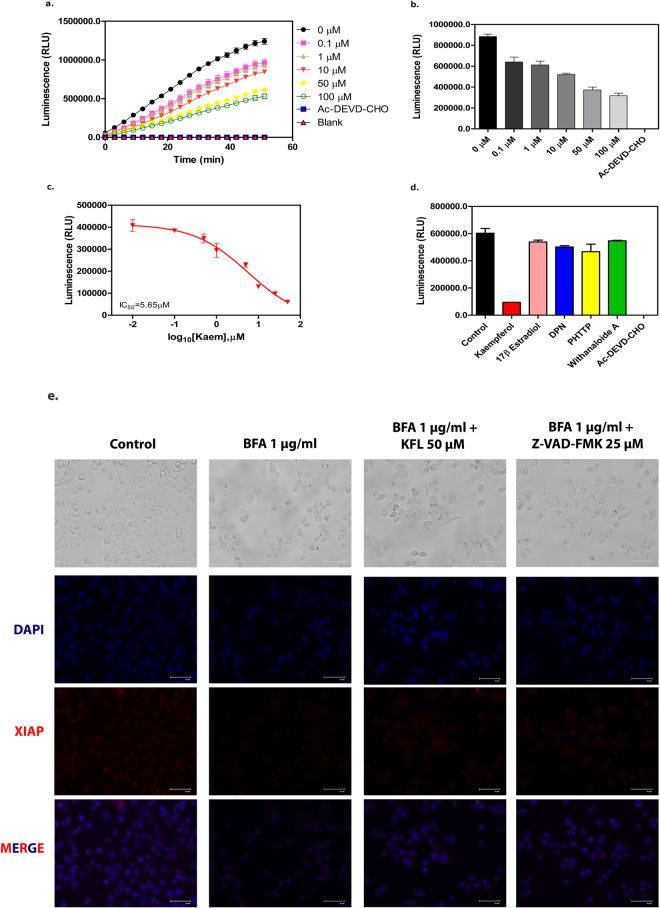
References
Publication types
MeSH terms
Substances
LinkOut - more resources
Full Text Sources
Other Literature Sources
Medical
Research Materials
Miscellaneous
