. 2017:2017:7435621.
doi: 10.1155/2017/7435621. Epub 2017 Dec 17.

HaCaT Cells as a Reliable In Vitro Differentiation Model to Dissect the Inflammatory/Repair Response of Human Keratinocytes

Affiliations

HaCaT Cells as a Reliable In Vitro Differentiation Model to Dissect the Inflammatory/Repair Response of Human Keratinocytes

Irma Colombo et al. Mediators Inflamm. 2017.

Abstract

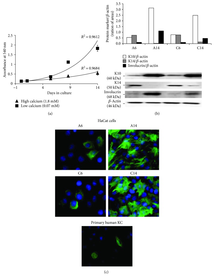
Cultured primary human keratinocytes are frequently employed for studies of immunological and inflammatory responses; however, interpretation of experimental data may be complicated by donor to donor variability, the relatively short culture lifetime, and variations between passages. To standardize the in vitro studies on keratinocytes, we investigated the use of HaCaT cells, a long-lived, spontaneously immortalized human keratinocyte line which is able to differentiate in vitro, as a suitable model to follow the release of inflammatory and repair mediators in response to TNFα or IL-1β. Different treatment conditions (presence or absence of serum) and differentiation stimuli (increase in cell density as a function of time in culture and elevation of extracellular calcium) were considered. ELISA and Multiplex measurement technologies were used to monitor the production of cytokines and chemokines. Taken together, the results highlight that Ca2+ concentration in the medium, cell density, and presence of serum influences at different levels the release of proinflammatory mediators by HaCaT cells. Moreover, HaCaT cells maintained in low Ca2+ medium and 80% confluent are similar to normal keratinocytes in terms of cytokine production suggesting that HaCaT cells may be a useful model to investigate anti-inflammatory interventions/therapies on skin diseases.


Figures

Figure 1
Figure 1
A flow chart with details of the experimental protocol performed on HaCaT cells.
Figure 2
Figure 2
Proliferation and differentiation of HaCaT cells in low (0.07 mM) and high (1.8 mM) Ca2+-containing medium. (a) Proliferation of HaCaT cells plated at the same density (1.0 × 104 cells/cm2) assessed by the MTT assay at 2, 6, 9, and 14 days of incubation. Values represent mean ± SD of three independent experiments. (b) Western blot analysis of the expression of keratinocyte (KC) differentiation markers (K10, K14, and involucrin) in HaCaT cells grown in low (a) and high (c) Ca2+-containing medium for 6 (A6 and C6) or 14 (A14 and C14) days. The relative intensities of band signals quantified by digital scanning densitometry are reported in the histogram; β-actin was used to normalize the results to protein content. This blot is a representative of three independent experiments. (c) Immunofluorescence staining of HaCaT cells, grown in low (A) and high (C) Ca2+-containing medium for 6 (A6 and C6) and 14 (A14 and C14) days, and of primary human KC, grown in low Ca2+-containing medium, with anti-K10 antibodies (magnification: 60x).
Figure 3
Figure 3
In vitro release of CXCL8/IL8, VEGF, and MMP-9 from HaCaT cells stimulated by TNFα during cell differentiation. The amount of CXCL8/IL8, VEGF, and MMP-9 was measured by ELISA in the supernatants of HaCaT cells plated at the same density (1.0 × 104 cells/cm2), grown in low (a) and high (C) Ca2+-containing medium for 6 (A6 and C6) or 14 (A14 and C14) days (white bars), and treated with 10 ng/ml TNFα for 6 or 24 hours (black bars), as indicated, in the absence (a) or the presence (b) of serum. The data are expressed as pg/106 cells, and values are the mean ± SD of at least three independent experiments in duplicate.
Figure 4
Figure 4
Bioplex multiplex system analysis of supernatants from HaCaT cells stimulated by TNFα during cell differentiation. The amount of chemokines (a), cytokines, and growth factors (b) was measured by Multiplex system technology in the supernatants of HaCaT cells plated at 1.0 × 104 cells/cm2, grown in low (a) and high (c) Ca2+-containing medium for 6 (A6 and C6) and 14 (A14 and C14) days (white bars), and treated with 10 ng/ml TNFα for 48 hours (black bars) in a serum-free medium. The data are expressed as pg/106 cells.
Figure 5
Figure 5
In vitro release of cytokines, chemokines, and growth factors from primary adult human keratinocytes stimulated by TNFα. The amount of chemokines (a), of cytokines, and of growth factors (b) was measured by Multiplex system technology in the supernatants of primary adult human keratinocytes (pKC) plated at the same density (8–10 × 104 cells/cm2) and grown in low Ca2+-containing medium (white bars). The treatment with 10 ng/ml TNFα was carried out for 48 hours in a serum-free medium when cells were 60–70% confluent (black bars). The data are expressed as pg/106 cells.

References

    1. Baroni A., Buommino E., De Gregorio V., Ruocco E., Ruocco V., Wolf R. Structure and function of the epidermis related to barrier properties. Clinics in Dermatology. 2012;30(3):257–262. doi: 10.1016/j.clindermatol.2011.08.007. - DOI - PubMed
    1. Hanel K. H., Cornelissen C., Luscher B., Baron J. M. Cytokines and the skin barrier. International Journal of Molecular Sciences. 2013;14(4):6720–6745. doi: 10.3390/ijms14046720. - DOI - PMC - PubMed
    1. Henry J., Toulza E., Hsu C. Y., et al. Update on the epidermal differentiation complex. Frontiers in Bioscience. 2012;17(1):1517–1532. doi: 10.2741/4001. - DOI - PubMed
    1. Proksch E., Brandner J. M., Jensen J. M. The skin: an indispensable barrier. Experimental Dermatology. 2008;17:1063–1072. doi: 10.1111/j.1600-0625.2008.00786.x. - DOI - PubMed
    1. Nestle F. O., Di Meglio P., Qin J. Z., Nickoloff B. J. Skin immune sentinels in health and disease. Nature Reviews Immunology. 2009;9(10):679–691. doi: 10.1038/nri2622. - DOI - PMC - PubMed

LinkOut - more resources