. 2017:2017:2490501.
doi: 10.1155/2017/2490501. Epub 2017 Dec 17.

Roles of Endoplasmic Reticulum Stress in NECA-Induced Cardioprotection against Ischemia/Reperfusion Injury

Affiliations

Roles of Endoplasmic Reticulum Stress in NECA-Induced Cardioprotection against Ischemia/Reperfusion Injury

Fengmei Xing et al. Oxid Med Cell Longev. 2017.

Abstract

Objective: This study aimed to investigate whether the nonselective A2 adenosine receptor agonist NECA induces cardioprotection against myocardial ischemia/reperfusion (I/R) injury via glycogen synthase kinase 3β (GSK-3β) and the mitochondrial permeability transition pore (mPTP) through inhibition of endoplasmic reticulum stress (ERS).

Methods and results: H9c2 cells were exposed to H2O2 for 20 minutes. NECA significantly prevented H2O2-induced TMRE fluorescence reduction, indicating that NECA inhibited the mPTP opening. NECA blocked H2O2-induced GSK-3β phosphorylation and GRP94 expression. NECA increased GSK-3β phosphorylation and decreased GRP94 expression, which were prevented by both ERS inductor 2-DG and PKG inhibitor KT5823, suggesting that NECA may induce cardioprotection through GSK-3β and cGMP/PKG via ERS. In isolated rat hearts, both NECA and the ERS inhibitor TUDCA decreased myocardial infarction, increased GSK-3β phosphorylation, and reversed GRP94 expression at reperfusion, suggesting that NECA protected the heart by inhibiting GSK-3β and ERS. Transmission electron microscopy showed that NECA and TUDCA reduced mitochondrial swelling and endoplasmic reticulum expansion, further supporting that NECA protected the heart by preventing the mPTP opening and ERS.

Conclusion: These data suggest that NECA prevents the mPTP opening through inactivation of GSK-3β via ERS inhibition. The cGMP/PKG signaling pathway is responsible for GSK-3β inactivation by NECA.


Figures

Figure 1
Figure 1
Confocal fluorescence images of TMRE in H9c2 cells. (a) NECA (0.01~10 μM) prevented oxidant-induced TMRE fluorescence (×400) reduction in a dose-dependent manner. (b) Summarized data for TMRE fluorescence intensity measured with confocal microscopy 20 min after exposure to H2O2 expressed as a percentage of baseline. Data are mean ± SD for 8 independent experiments performed in duplicate. P < 0.05 versus the control group, #P < 0.05 versus the H2O2 group.
Figure 2
Figure 2
The effect of NECA on GSK-3β at Ser9 phosphorylation and GRP94 protein expression in H9c2 cells. (a) NECA (0.01–10 μM) increased GSK-3β phosphorylation and decreased GRP94 expression in a dose-dependent manner. (b) Data are mean ± SD for 8 independent experiments performed in duplicate. P < 0.05 compared to the control group.
Figure 3
Figure 3
The effect of NECA on GSK-3β at Ser9 phosphorylation and GRP94 protein expression in H2O2-treated H9c2 cells. (a) H2O2 (800 μM) increased GSK-3β phosphorylation and decreased GRP94 expression, the effect that was blocked by NECA (0.1 μM). (b) Data are mean ± SD for 8 independent experiments performed in duplicate. P < 0.05 compared to the control group; #P < 0.05 compared to NECA.
Figure 4
Figure 4
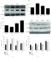
NECA inactivates GSK-3β via inhibiting ERS in the heart. (a) NECA (0.1 μM) increased GSK-3β phosphorylation and decreased GRP94 expression; the effect was reversed by the specific inducer of ERS 2-DG (20 μM). (b, c) Data are mean ± SD for 8 independent experiments performed in duplicate. P < 0.05 compared to the control group; #P < 0.05 compared to NECA. (d) I/R increased GSK-3β phosphorylation and decreased GRP94 expression after reperfusion 10 min, 30 min, 60 min, and 120 min in the rat heart, the effect that was reversed by NECA (0.1 μM) and the specific inhibitor of ERS TUDCA (30 μM). (e, f) Data are mean ± SD for 8 independent experiments performed in duplicate. P < 0.05 compared to the I/R group.
Figure 5
Figure 5
NECA induces cardioprotection by inactivating GSK-3β via ERS and cGMP/PKG signaling pathway. (a) NECA (0.1 μM) increased GSK-3β and VASP phosphorylation and decreased GRP94 expression, the effect that was reversed by the specific inducer of PKG KT5823 (1 μM). (b, c, d) Data are mean ± SD for 8 independent experiments performed in duplicate. P < 0.05 compared to the control group; #P < 0.05 compared to NECA.
Figure 6
Figure 6
The effects of NECA on infarct size reduction following 30 min of occlusion and 120 min of reperfusion (I/R: n = 5, NECA: n = 4, TUDCA: n = 4). P < 0.05 versus I/R.
Figure 7
Figure 7
Structural changes of the rat LV tissues as assessed via transmission electron microscopy. Original magnification ×15,000.
Figure 8
Figure 8
Signaling pathway leading to the cardioprotective effect of NECA.

References

    1. Lee C. H., Low A., Tai B. C., et al. Pretreatment with intracoronary adenosine reduces the incidence of myonecrosis after non-urgent percutaneous coronary intervention: a prospective randomized study. European Heart Journal. 2007;28(1):19–25. doi: 10.1093/eurheartj/ehl411. - DOI - PubMed
    1. Takahama H., Minamino T., Asanuma H., et al. Prolonged targeting of ischemic/reperfused myocardium by liposomal adenosine augments cardioprotection in rats. Journal of the American College of Cardiology. 2009;53(8):709–717. doi: 10.1016/j.jacc.2008.11.014. - DOI - PubMed
    1. Tian Y., Piras B. A., Kron I. L., French B. A., Yang Z. Adenosine 2B receptor activation reduces myocardial reperfusion injury by promoting anti-inflammatory macrophages differentiation via PI3K/Akt pathway. Oxidative Medicine and Cellular Longevity. 2015;2015:8. doi: 10.1155/2015/585297.585297 - DOI - PMC - PubMed
    1. Prystowsky E. N., Niazi I., Curtis A. B., et al. Termination of paroxysmal supraventricular tachycardia by tecadenoson (CVT-510), a novel A1-adenosine receptor agonist. Journal of the American College of Cardiology. 2003;42(6):1098–1102. doi: 10.1016/S0735-1097(03)00987-2. - DOI - PubMed
    1. Xi L., Das A., Zhao Z. Q., Merino V. F., Bader M., Kukreja R. C. Loss of myocardial ischemic postconditioning in adenosine A1 and bradykinin B2 receptors gene knockout mice. Circulation. 2008;118(14) Supplement 1:S32–S37. doi: 10.1161/CIRCULATIONAHA.107.752865. - DOI - PMC - PubMed

MeSH terms

Substances

LinkOut - more resources