Review
. 2018 Feb 1;7(2):29-45.
doi: 10.1089/wound.2016.0696.

Cutaneous Scarring: Basic Science, Current Treatments, and Future Directions

Affiliations
Review

Cutaneous Scarring: Basic Science, Current Treatments, and Future Directions

Clement D Marshall et al. Adv Wound Care (New Rochelle). .

Abstract

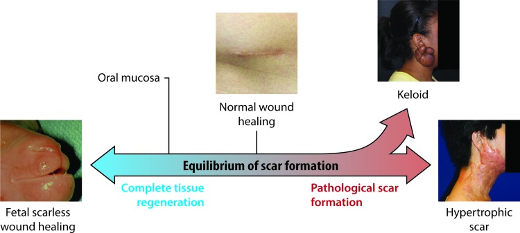
Significance: Scarring of the skin from burns, surgery, and injury constitutes a major burden on the healthcare system. Patients affected by major scars, particularly children, suffer from long-term functional and psychological problems. Recent Advances: Scarring in humans is the end result of the wound healing process, which has evolved to rapidly repair injuries. Wound healing and scar formation are well described on the cellular and molecular levels, but truly effective molecular or cell-based antiscarring treatments still do not exist. Recent discoveries have clarified the role of skin stem cells and fibroblasts in the regeneration of injuries and formation of scar. Critical Issues: It will be important to show that new advances in the stem cell and fibroblast biology of scarring can be translated into therapies that prevent and reduce scarring in humans without major side effects. Future Directions: Novel therapies involving the use of purified human cells as well as agents that target specific cells and modulate the immune response to injury are currently undergoing testing. In the basic science realm, researchers continue to refine our understanding of the role that particular cell types play in the development of scar.


Figures

None
Michael T. Longaker, MD, MBA
<b>Figure 1.</b>
Figure 1.
Spectrum of cutaneous scar formation. Illustration of the various possible endpoints of scar formation. Left, a fetal lamb that healed a prior lip wound with no scar whatsoever. Middle, a normal and well-healed appendectomy scar. Upper right, a keloid scar in the classic ear lobe location. Lower right, hypertrophic scar resulting from a scald burn. To see this illustration in color, the reader is referred to the web version of this article at www.liebertpub.com/wound
<b>Figure 2.</b>
Figure 2.
Dermal biopsy locations from healthy controls and keloid patients with corresponding histology. (A) Transverse view of biopsy locations from normal dermal scar tissue and adjacent normal dermal (nonwounded) skin from which in vitro primary cell cultures were subsequently established. (B) Transverse view of marginal perilesional and reticular dermal intralesional biopsy sites from the keloid scar. (C) Cross section of keloid scar indicating depth of perilesional and intralesional biopsies. (D) Representative H&E staining of tissue section from normal skin indicating organized wavy deposition of collagen (blue arrows). (E) Representative H&E staining of tissue section from a normal scar. (F) Representative H&E staining of a perilesional keloid tissue section indicating a thickened EP with increased cell infiltration (yellow arrow) and deposition of hyalinized collagen bundles in the RD (black arrow). (G) Representative H&E staining of an intralesional keloid tissue section indicating thick compact hyalinized collagen bundle deposition in the RD (black arrow). All the H&E micrographs (DG) were taken at 200 magnifications. Reprinted with permission from Ashcroft et al. EP, epidermis; PD, papillary dermis; H&E, hematoxylin and eosin; RD, reticular dermis. To see this illustration in color, the reader is referred to the web version of this article at www.liebertpub.com/wound
<b>Figure 3.</b>
Figure 3.
Transformation of the fibroblast to a myofibroblast. PDGF and TGF-β signaling promotes transformation of fibroblasts to myofibroblasts, which contribute to wound contraction and are characterized by expression of α-smooth muscle actin. The contractile force provided by myofibroblasts can cause wound edges to move toward each other by 0.75 mm per day. PDGF, platelet-derived growth factor; TGF-β, transforming growth factor-beta. To see this illustration in color, the reader is referred to the web version of this article at www.liebertpub.com/wound
<b>Figure 4.</b>
Figure 4.
Classical stages of wound healing with key cellular players. Platelets are the first agents to arrive and contribute to hemostasis. During the inflammatory phase, neutrophils clean up the wound by phagocytosing debris and bacteria. Macrophages arrive after neutrophils and reside for longer, performing phagocytosis as well. During the proliferative phase, keratinocytes migrate onto the wound surface to re-epithelialize the wound, while endothelial cells reconstruct blood vessels. Beginning in the proliferative phase and extending indefinitely in the remodeling phase, fibroblasts lay down collagen and contribute to scar formation. To see this illustration in color, the reader is referred to the web version of this article at www.liebertpub.com/wound
<b>Figure 5.</b>
Figure 5.
Histology of the scarless fetal wound. E16 fetal wounds (hematoxylin and eosin stain). Black arrows indicate India ink tattoo made at the time of wounding to demonstrate scarless wound location. Healed wounds (above, left and below, left) at 72 h (100×). The epidermal appendage (developing hair follicles) pattern shows numerous appendages directly in the healed wound. Magnified views of the same wounds (above, right and below, right) showing epidermal appendages (open arrows) within the wound site (200×). No inflammatory infiltrate is present. Reprinted with permission from Beanes et al. To see this illustration in color, the reader is referred to the web version of this article at www.liebertpub.com/wound
<b>Figure 6.</b>
Figure 6.
Differences between scarless and scarring wound healing processes. At a cellular and molecular level, there are several differences between fetal and adult wound healing that may contribute to the scarless versus scarring phenotype. To see this illustration in color, the reader is referred to the web version of this article at www.liebertpub.com/wound
<b>Figure 7.</b>
Figure 7.
Sources of stem cells for skin regeneration. Stem cells from several locations may contribute to the regeneration of skin after injury. Epidermal stem cells residing in the basal layer of the interfollicular dermis repopulate the epidermis under normal condition and after injury., Cells of the dermal papilla can direct the formation of new hair follicles in uninjured skin. Cells from the hair follicle bulge region repopulate the hair follicle itself normally and can help to repopulate the epidermis after injury. The hair follicle junctional zone contains cells with distinct lineages that contribute to hair follicle and epidermal regeneration.,, Cells of the sebaceous gland primarily regenerate the gland itself, while cells of the eccrine sweat gland duct may contribute to epidermal repair after injury. Finally, mesenchymal stem cells arising from the bone marrow and circulating in blood may migrate into injured skin and assist in regeneration. To see this illustration in color, the reader is referred to the web version of this article at www.liebertpub.com/wound
<b>Figure 8.</b>
Figure 8.
Illustration of normal versus scarred skin. Collagen in normal skin is arranged in a basket-weave pattern, whereas scar collagen is arranged in parallel fibers. This, in addition to the lack of elastic fibers, contributes to the stiffness of scar tissue. Also, there is a notable absence of dermal appendages, including hair follicles, sebaceous glands, and sweat glands, in scarred skin. Finally, the basement membrane separating epidermis from dermis is flatter in scarred skin and does not contain rete pegs that normally extend down into the dermis. To see this illustration in color, the reader is referred to the web version of this article at www.liebertpub.com/wound
<b>Figure 9.</b>
Figure 9.
Histology of normal and scarred skin. Trichrome stain of uninjured (a) and scarred (b) adult mouse skin. Hair follicles are plentiful in normal skin but absent in scarred skin. The scarred dermis is a relatively acellular expanse of collagen. Scale bar, 100 μm. To see this illustration in color, the reader is referred to the web version of this article at www.liebertpub.com/wound
<b>Figure 10.</b>
Figure 10.
Scar potential. A specific fibroblast lineage in dorsal skin increasingly populates skin with age and is responsible for extracellular matrix production in multiple developmental and pathophysiological scenarios. Reprinted with permission from Sennett and Rendl. To see this illustration in color, the reader is referred to the web version of this article at www.liebertpub.com/wound

References

    1. Sheridan RL, et al. Long-term outcome of children surviving massive burns. JAMA 2000;283:69–73 - PubMed
    1. Esselman PC. Burn rehabilitation: an overview. Arch Phys Med Rehabil 2007;88:S3–S6 - PubMed
    1. Robert R, et al. Disfiguring burn scars and adolescent self-esteem. Burns 1999;25:581–585 - PubMed
    1. Van Loey NE, Van Son MJ. Psychopathology and psychological problems in patients with burn scars: epidemiology and management. Am J Clin Dermatol 2003;4:245–272 - PubMed
    1. Thomas CR, et al. Personality disorders in young adult survivors of pediatric burn injury. J Pers Disord 2012;26:255–266 - PubMed