. 2018 Jun;169(2):381-390.
doi: 10.1007/s10549-018-4685-2. Epub 2018 Feb 1.

A novel patient-derived xenograft model for claudin-low triple-negative breast cancer

Affiliations

A novel patient-derived xenograft model for claudin-low triple-negative breast cancer

Margarite D Matossian et al. Breast Cancer Res Treat. 2018 Jun.

Abstract

Background: Triple-negative breast cancer (TNBC) subtypes are clinically aggressive and cannot be treated with targeted therapeutics commonly used in other breast cancer subtypes. The claudin-low (CL) molecular subtype of TNBC has high rates of metastases, chemoresistance and recurrence. There exists an urgent need to identify novel therapeutic targets in TNBC; however, existing models utilized in target discovery research are limited. Patient-derived xenograft (PDX) models have emerged as superior models for target discovery experiments because they recapitulate features of patient tumors that are limited by cell-line derived xenograft methods.

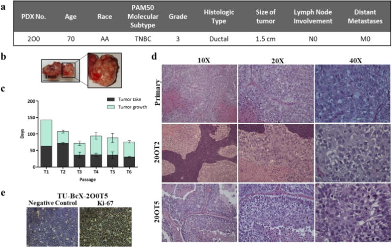
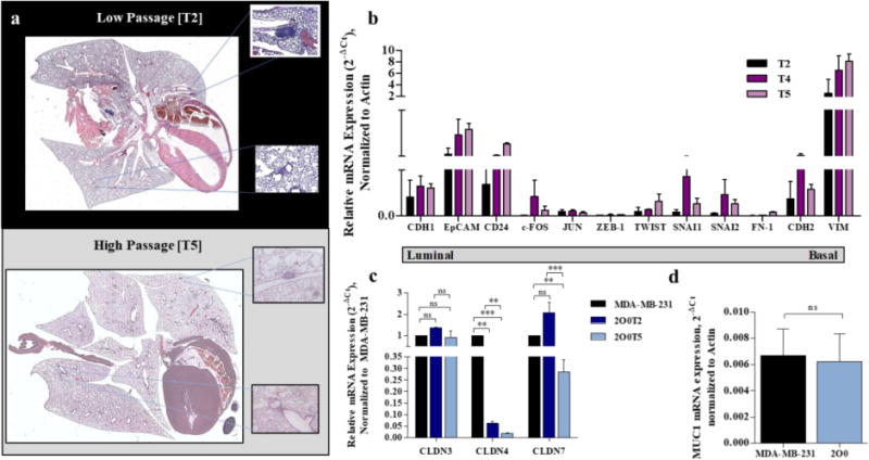
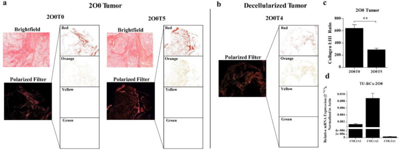
Methods: We utilize immunohistochemistry, qRT-PCR and Western Blot to visualize tumor architecture, cellular composition, genomic and protein expressions of a new CL-TNBC PDX model (TU-BcX-2O0). We utilize tissue decellularization techniques to examine extracellular matrix composition of TU-BcX-2O0.

Results: Our laboratory successfully established a TNBC PDX tumor, TU-BCX-2O0, which represents a CL-TNBC subtype and maintains this phenotype throughout subsequent passaging. We dissected TU-BCx-2O0 to examine aspects of this complex tumor that can be targeted by developing therapeutics, including the whole and intact breast tumor, specific cell populations within the tumor, and the extracellular matrix.

Conclusions: Here, we characterize a claudin-low TNBC patient-derived xenograft model that can be utilized for therapeutic research studies.

Keywords: Claudin-low; Collagen; Decellularized tumor scaffold; Extracellular matrix; Mesenchymal; Patient-derived xenograft; Triple-negative breast cancer.


Conflict of interest statement

Conflict of Interest Statement

The authors declare that they have no conflict of interest.

Figures

Figure 1
Figure 1. TU-BcX-2O0, had a distinct gross appearance and tumor growth pattern and has a consistent cytology throughout passages
(a) Patient data from TU-BcX-200 shows it is categorized as TNBC based on the PAM50 molecular subtype without lymph node (N0) or distant metastases (M0) involvement at the time of tumor resection. (b) Gross appearance of TU-BcX-2O0 was consistent throughout consecutive passages. The tumor had distinct pockets filled with a pustulate-like substance that was liquid when dissected. (c) Tumor growth patterns of TU-BcX-2O0 after initial engraftment into the mfp of SCID/Beige mice. Tumor growth indicates the number of days from when the tumor first became palpable to when tumors were extracted (at a final volume of 750–1000 mm3). (d) The primary specimen (obtained post-op, not yet engrafted into SCID/Beige mice) histology was compared to lower (T2) and higher passage (T5) PDX tumors. H & E stained images were captured at 10×, 20× and 40×. (e) Proliferation rate determined by Ki-67 immunohistochemistry staining of TU-BcX-2O0T5 PDX tumor compared to negative control.
Figure 2
Figure 2. TU-BcX-2O0 metastasizes to the lungs and expresses mesenchymal markers
(a) At both lower and higher passages, TU-BcX-2O0 metastasizes to the lungs, although there are fewer lesions in higher passages. Hematoxylin and Eosin stained and imaged. Examples of specific lesions are represented by the boxed areas. (b) qRT-PCR panel of luminal (CDH1, EpCAM, CD24) and basal (c-FOS, JUN, ZEB-1, TWIST, SNAI1, SNAI2, FN-1, CDH2, VIM) gene expressions. Gene expressions in various passages ranging from low (T2) to high (T5) were compared. All data was obtained using qRT-PCR in triplicate and normalized to actin. Error bars represent SEM and significantly different * p < 0.05, *** p < 0.001. (c) The following mRNA baseline expressions of genes enriched or downregulated in the claudin-low TNBC subtype from TU-BcX-2O0 is compared to the claudin-low TNBC cell line, MDA-MB-231. (a) CLDN3, 4 and 7 gene expressions, defining the CL-TNBC subtype, of MDA-MB-231 and 2O0 tumor. (d) 2O0 has comparable MUC1 mRNA expression, a gene associated with the CL subtype, compared to MDA-MB-231. Error bars represent SEM and significantly different * p < 0.05, *** p < 0.001.
Figure 3
Figure 3. Collagen composition of TU-BcX-2O0 was not altered throughout consecutive passages
(a) Picosirius Red staining (top panel) was utilized to highlight collagen fibers, and subsequent imaging of the stained slides under filtered light (bottom panels) revealed specific collagen I and collagen III composition of tumors throughout low and higher passages. Also shown are extracted images of the four polarized filters utilized to identify the specific collagens displayed. Red and orange filter indicate collagen I; yellow and green indicate presence of collagen III. (b) Similar architecture is shown when the tumor was decellularized. There is an abundance of collagen I compared to collagen III. (c) Quantification of collagen staining demonstrates collagen I:III ratio was larger in lower passage TU-BcX-2O0 compared to higher passage, although collagen III staining remained low (p=0.0093). (d) Additionally, in decellularized TU-BcX-2O0 tumor, there was a significant variation of collagen I:III ratio between passages (p=0.002), although collagen I remained dominant. Quantification was performed in triplicate and ratio is shown as Red AF+Orange AF/Yellow AF + Green AF; AF stands for Area Fraction (%). n = 3 for TU-BcX-2O0T3 decellularized and TU-BcX-2O0T5; n = 4 for 2O0T0 and n = 5 for TU-BcX-2O0T4 decellularized TU-BcX-2O0.

References

    1. Alteri R, Bertaut T, Brinton LA, Fedewa S, Freedman RA, Gansler T, et al. Breast cancer facts and figures 2015–2016. Atlanta: American Cancer Society Inc; 2015.
    1. Blows FM, Driver KE, Schmidt MK, Broeks A, van Leeuwen FE, Wesseling J. Subtyping of breast cancer by immunohistochemistry to investigate a relationship between subtype and short and long term survival: A collaborative analysis of data for 10,159 cases from 12 studies. PLOS Med. 2010 doi: 10.1371/journal.pmed.1000279. - DOI - PMC - PubMed
    1. Crown J, O’Shaughnessy J, Gullo G. Emerging targeted therapies in triple-negative breast cancer. Annals Oncol. 2012;23(S6):vi56–65. - PubMed
    1. Stevens KN, Vachon CM, Couch FJ. Genetic susceptibility to triple-negative breast cancer. Cancer Res. 2013;73(7):2025–30. - PMC - PubMed
    1. Engebraaten O, Vollan HKM, Borresen-Dale A-L. Triple-negative breast cancer and the need for new therapeutic targets. Am J Pathol. 2013;183(4):1064–74. - PubMed

LinkOut - more resources