. 2018 Apr;46(4):1358-1369.
doi: 10.1177/0300060517752022. Epub 2018 Feb 2.

Antioxidant activity and apoptotic induction as mechanisms of action of Withania somnifera (Ashwagandha) against a hepatocellular carcinoma cell line

Affiliations

Antioxidant activity and apoptotic induction as mechanisms of action of Withania somnifera (Ashwagandha) against a hepatocellular carcinoma cell line

Wafaa Ahmed et al. J Int Med Res. 2018 Apr.

Abstract

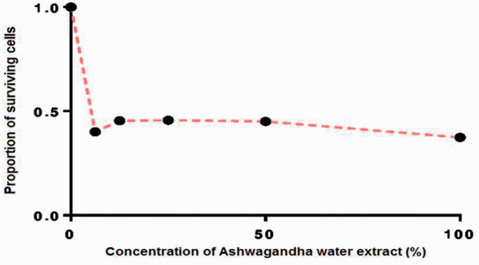
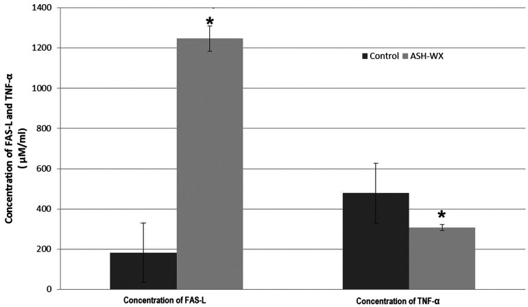
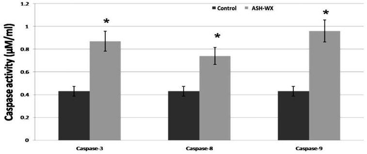
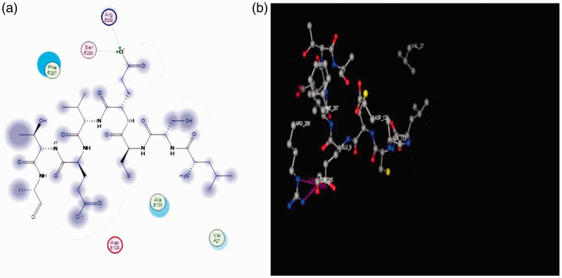
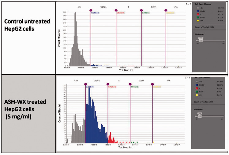
Objective To evaluate the antioxidant and apoptotic inductive effects of Withania somnifera (Ashwagandha) leaf extract against a hepatocellular carcinoma cell line. Methods After treating HepG2cells with Ashwagandha water extract (ASH-WX; 6.25 mg/ml-100 mg/ml), cell proliferation was assessed using a 3-(4,5-dimethylthiazol-2-yl)-2,5-diphenyltetrazolium bromide (MTT) assay. Antioxidant activities (total antioxidant, glutathione S-transferase and glutathione reductase), Fas-ligand level, tumour necrosis factor-α (TNF-α) level and caspase-3, -8, and -9 activities were measured. Molecular modelling assessed the binding-free energies of Ashwagandha in the cyclin D1 receptor. Results The MTT assay demonstrated increased cytotoxicity following treatment of HepG2 cells with ASH-WX compared with control untreated cells and theIC50was 5% (approximately 5.0 mg/ml). Antioxidant activities, Fas-ligand levels and caspase-3, -8 and -9 activities significantly increased, while TNF-α level significantly decreased following ASH-WX treatment compared with control untreated cells. Molecular docking analysis revealed a good prediction of binding between cyclin D1 and Ashwagandha. There was significant accumulation of ASH-WX-treated HepG2cells in the G0/G1 and G2/M phases compared with the control untreated cells. Conclusion Ashwagandha could be a powerful antioxidant and a promising anticancer agent against HCC.

Keywords: Ashwagandha; HepG2; antioxidants; apoptosis; cytotoxicity.


Figures

Figure 1.
Figure 1.
Growth response curve for HepG2 cells treated with a range of concentrations of Ashwagandha water extract (6.25mg/ml–100 mg/ml) at 37 °C in a 5% CO2 incubator for 24 h calculated using GraphPad Prism 7 software. The IC50 value was 5.0 mg/ml.
Figure 2.
Figure 2.
Representative photomicrographs showing the viability of HepG2 cells treated with a range of concentrations of Ashwagandha water extract (ASH-WX) at 37 °C in a 5% CO2 incubator for 24 h. (a) Control untreated cells; (b) cells treated with 100 mg/ml ASH-WX; (c) cells treated with 50 mg/ml ASH-WX; (d) cells treated with 25 mg/ml ASH-WX; (e) cells treated with 12.5 mg/ml ASH-WX; and (f) cells treated with 6.25 mg/ml ASH-WX. HepG2 cells incubated in all concentrations showed signs of marked shrinkage and accumulation of dead cells compared with the control untreated cells. The higher the concentration of ASH-WX, the higher the percentage of dead cells and shrinkage of cells. Scale bar 20 µm. The colour version of this figure is available at: http://imr.sagepub.com.
Figure 3.
Figure 3.
The concentration of Fas-ligand (FAS-L) and tumour necrosis factor-α (TNF-α) in HepG2 cells treated with the IC50 concentration (5.0 mg/ml) of Ashwagandha water extract (ASH-WX) at 37 °C in a 5% CO2 incubator for 48 h. Data presented as mean ± SD. *P < 0.05 compared with the control untreated cells; Mann–Whitney U-test.
Figure 4.
Figure 4.
The activity of caspase-3, caspase-8 and caspase-9 in HepG2 cells treated with the IC50 concentration (5.0 mg/ml) of Ashwagandha water extract (ASH-WX) at 37 °C in a 5% CO2 incubator for 48 h. Data presented as mean ± SD. *P < 0.05 compared with the control untreated cells; Mann–Whitney U-test.
Figure 5.
Figure 5.
Ligand interaction and binding model analysis of Ashwagandha with the cyclin D 1 receptor showing the pharmacophore model (a) and docking drug model (b). Ashwagandha exhibited two hydrogen bonds with the amino acids in cyclin D1: arginine β 256 and serine β 258 (hydrogen bonds shown in green). Arg B256, arginine β 256; Ser B258, serine β 258; Phe B287, phenylalanine β 287; Ala B1, alanine β1; Val A27, valine α 27; Arg B129, arginine β 129. The colour version of this figure is available at: http://imr.sagepub.com.
Figure 6.
Figure 6.
Cell cycle analysis of HepG2 cells treated with the IC50 concentration (5.0 mg/ml) of Ashwagandha water extract (ADH-WX) at 37 °C in a 5% CO2 incubator for 24 h analysed using a Cytell™ cell imaging system. The colour version of this figure is available at: http://imr.sagepub.com.

References

    1. Sever R andBrugge JS.. Signal transduction in cancer. Cold Spring Harb Perspect Med 2015; 5. Epub ahead of print 1 April 2015. DOI: 10.1101/cshperspect.a006098 - PMC - PubMed
    1. Hanahan D andWeinberg RA.. Hallmarks of cancer: the next generation. Cell 2011; 144: 646–674. - PubMed
    1. Fouad YA andAanei C.. Revisiting the hallmarks of cancer. Am J Cancer Res 2017; 7: 1016–1036. - PMC - PubMed
    1. Adjiri A. Identifying and targeting the cause of cancer is needed to cure cancer. Oncol Ther 2016; 4: 17–33. - PMC - PubMed
    1. Charames GS andBapat B.. Genomic instability and cancer. Curr Mol Med 2003; 3: 589–596. - PubMed

MeSH terms