MRI-only treatment planning: benefits and challenges
- PMID: 29393071
- PMCID: PMC5886006
- DOI: 10.1088/1361-6560/aaaca4
MRI-only treatment planning: benefits and challenges
Abstract
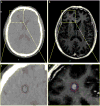
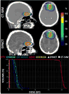
Over the past decade, the application of magnetic resonance imaging (MRI) has increased, and there is growing evidence to suggest that improvements in the accuracy of target delineation in MRI-guided radiation therapy may improve clinical outcomes in a variety of cancer types. However, some considerations should be recognized including patient motion during image acquisition and geometric accuracy of images. Moreover, MR-compatible immobilization devices need to be used when acquiring images in the treatment position while minimizing patient motion during the scan time. Finally, synthetic CT images (i.e. electron density maps) and digitally reconstructed radiograph images should be generated from MRI images for dose calculation and image guidance prior to treatment. A short review of the concepts and techniques that have been developed for implementation of MRI-only workflows in radiation therapy is provided in this document.
Figures


References
-
- Andreasen D, Van Leemput K, Edmund JM. A patch-based pseudo-CT approach for MRI-only radiotherapy in the pelvis. Med Phys. 2016;43:4742. - PubMed
-
- Arias-Mendoza F, Payne GS, Zakian K, Stubbs M, O’Connor OA, Mojahed H, Smith MR, Schwarz AJ, Shukla-Dave A, Howe F, Poptani H, Lee S-C, Pettengel R, Schuster SJ, Cunningham D, Heerschap A, Glickson JD, Griffiths JR, Koutcher JA, Leach MO, Brown TR. Noninvasive phosphorus magnetic resonance spectroscopic imaging predicts outcome to first-line chemotherapy in newly diagnosed patients with diffuse large B-cell lymphoma. Academic radiology. 2013;20:1122–9. - PMC - PubMed
-
- Baldwin LN, Wachowicz K, Thomas SD, Rivest R, Fallone BG. Characterization, prediction, and correction of geometric distortion in 3 T MR images. Med Phys. 2007a;34:388. - PubMed
-
- Baldwin LN, Wachowicz K, Thomas SD, Rivest R, Fallone BG. Characterization, prediction, and correction of geometric distortion in 3 T MR images. Medical physics. 2007b;34:388–99. - PubMed
-
- Baldwin LN, Wachowicz K, Thomas SD, Rivest R, Fallone BG. Characterization, prediction, and correction of geometric distortion in 3 T MR images. Medical physics. 2007c;34:388–99. - PubMed
Publication types
MeSH terms
Grants and funding
LinkOut - more resources
Full Text Sources
Other Literature Sources
Medical
Miscellaneous
